Invertebrates are animals without a backbone (spine). Animals are multicellular, heterotrophic organisms forming the Kingdom Metazoa. In contrast to the autotrophic algae and plants, animals are not capable of producing their own food via photosynthesis, so they depend on eating others. Most, but not all animals are motile, i.e. capable of motion. Invertebrates contain 97 % of all animal species. Based on their body construction they can be grouped into 35 phyla, all represented in the marine environment. Invertebrates that we encounter most frequently belong to the following phyla.
- Sponges
- Cnidarians
- Comb Jellies
- Worms
- Molluscs
- Crustaceans
- Bryozoans
- Enchinoderms
- Tunicates
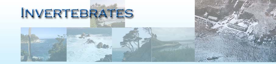
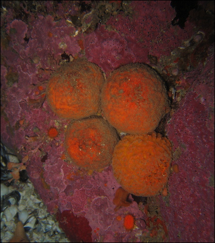
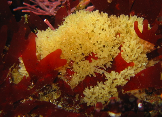
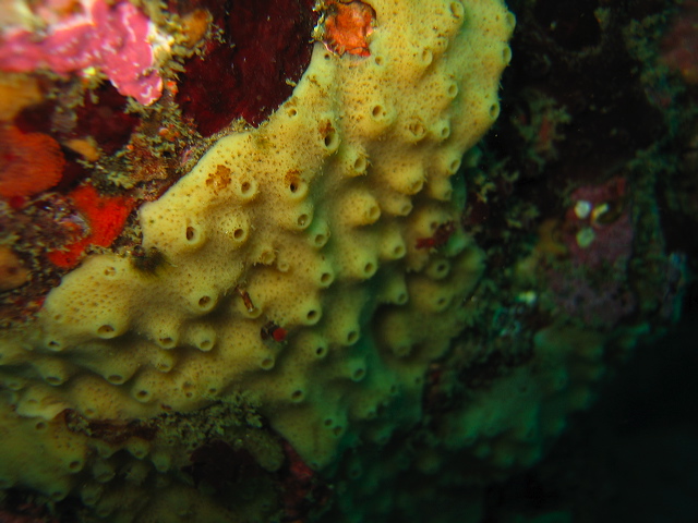
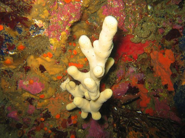
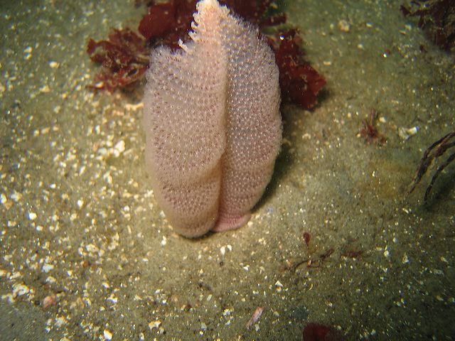
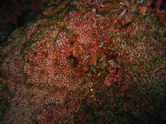
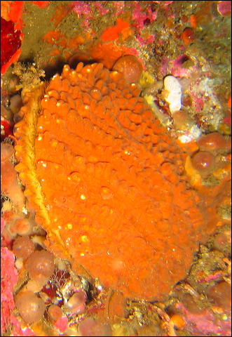
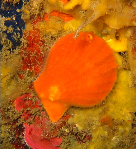
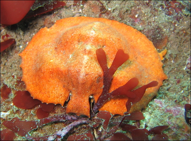
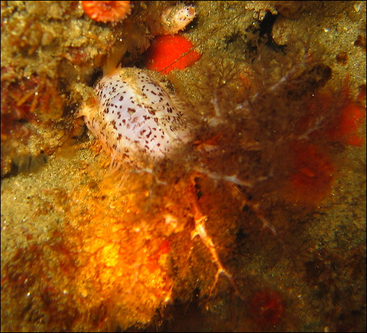
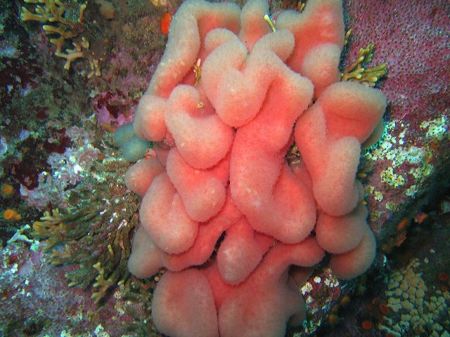
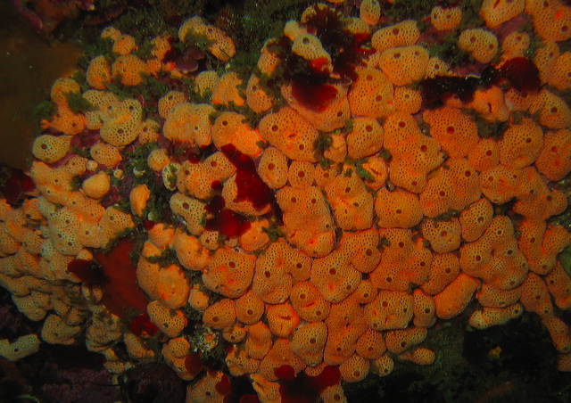
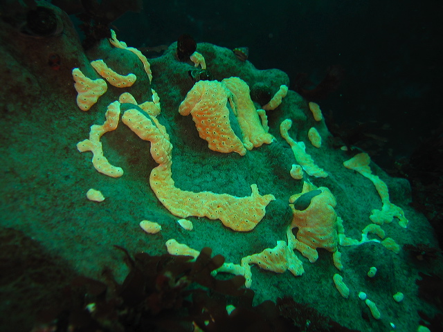
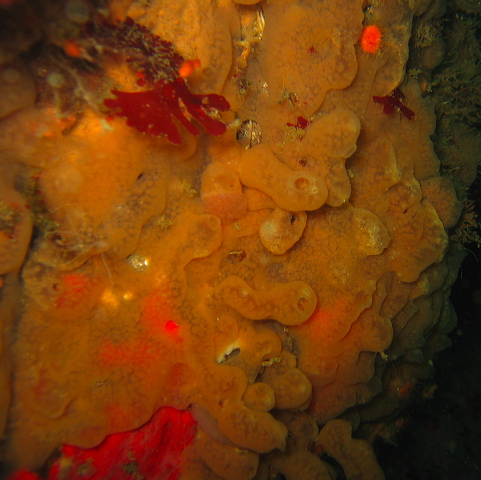
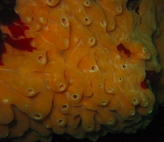
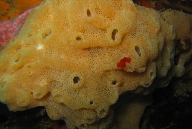
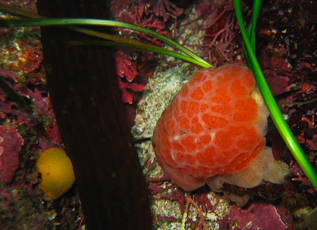
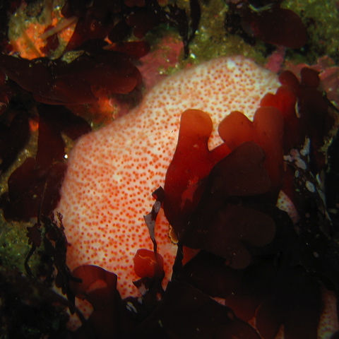
The humble Sponge is an often overlooked and underappreciated marine organism. This ancient group has been around for 600 million years. Porifera, the Latin name of the phylum, means “pore bearer”, referring to the visibly apparent openings on the surface. Sponges come in a variety of colors and shapes. The bright colors, yellow, orange, red, even blue are mainly due to symbiotic microorganisms. Some species grow as thick layers on hard surfaces, some form interesting shapes, resembling a tennis ball, an elephant ear, or a small vase. These are sessile organisms, attached permanently to a rock or other substrate. Sponges can be easily confused with tunicates, but the spongy texture, the dull, felt like surface and some conspicuous pores help in distinguishing between the two phyla.
Sponges represent the link between unicellular and multicellular organisms. In a major evolutionary step single flagellated cells started to communicate and collaborate with each other, this allowed an organism to grow beyond the limits of a single cell. These simple multicellular organisms, also called parazoa, are organized at the cell level. Lacking tissues and organs they are merely an aggregation of specialized cells. Sponges are the only multicellular animals organized in this manner. Despite their large size they function in a similar manner to microscopic unicellular organisms, all activity, like digestion and respiration, are performed at the cell level. Sponges have no sensory organs, nerves, or even muscles. Simplicity, however, has its advantages: sponge cells are very plastic, capable of changing roles and reorganizing themselves. If cells are separated by putting a sponge through a sieve, the cells start to aggregate and form new sponges. If the sponge cells come from different species they are capable of self recognition, so they reaggregate into separate species. Although many invertebrates are capable of regenerating lost body parts, cell reorganization is unique to sponges.
As active hunting is out of the question for the sessile sponge, to feed themselves they pump enormous amounts of water (pumping 200 gallon of water produces only an ounce of food) through their bodies and filter out small organic particles, bacteria, microscopic algae. They are even capable of absorbing dissolved organic matter. The water enters via many tiny pores, called ostia, then circulates through the canals and chambers crisscrossing the entire sponge body. The simple process ensures that food is delivered to each individual cell. The pumping effect is achieved with the help of the most typical sponge cells, the choanocytes, lining the canals. Each has a single, beating flagella, that creates the water current and a collar that traps food particles. These choanocyte cells are of great importance to evolutionary biologists as they resemble the choanoflagellates, the closest living unicellular relative to animals, a modern form of the last unicellular ancestor of animals. Finally, the water is pumped out via the osculum, a large opening at the top. This aquiferous system is important not only in food gathering but also in gas exchange, expelling waste products and sexual reproduction. The food is passed to the ameboid cells, where it gets digested and the resulting nutirents are transported to all other cells. These archaeocytes are totipotent cells that can take on various tasks, as needed, for example, transform into eggs or sperm, or secret spicules. If distressed, the sponge can shrink the size of its pores (ostia), even stopping the water flow with the help of the contractile cells lining the openings.
The structural support of sponges is provided by tiny siliceous or calcareous elements, called spicules, or by a protein fiber, called spongin. The spicules are the basis of sponge taxonomy, i.e. how sponges are arranged into smaller hierarchical groups. Most sponges we encounter belong to the class Demospongiae with silicone dioxide spicules. Some of the small white sponges belong to the class Calcarea.
Sponges use various methods for reproduction. Most are hermaphrodites, producing both eggs and sperms. In most cases only the sperms are released into the water column, enter the body of another sponge where internal fertilization takes place. As with many invertebrates life begins with a larval form, which after a brief planktonic period settles down and develops into adult. Sponges can also proliferate asexually, growing new individuals mainly by budding. Some smaller sponges are annual, but the larger ones may live up to 100 years.
As sponges lack physical defense mechanism, a shell or spines, they produce a wide variety of chemicals to deter predators and competitors, or fight infection and fouling. These compounds are of pharmacological importance because they possess antibacterial, antiviral, anti-inflammatory, and antitumor properties. In fact, the first approved marine drug is based on a natural compound produced by the humble sponge. Despite this chemical warfare sponges do have predators, snails, nudibranchs, even some fish become immune to the toxins.
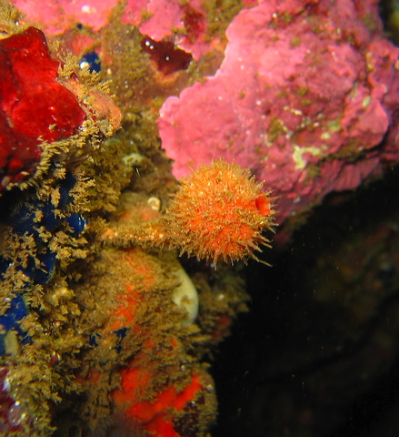
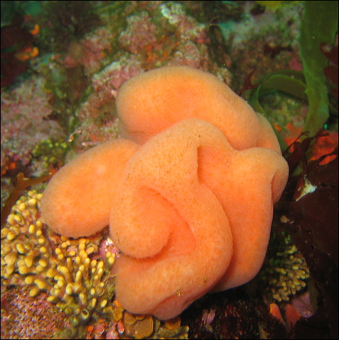
Tethya californiana – Orange Puffball Sponge
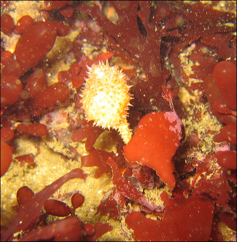
Craniella arb – Gray Puffball Sponge
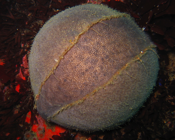
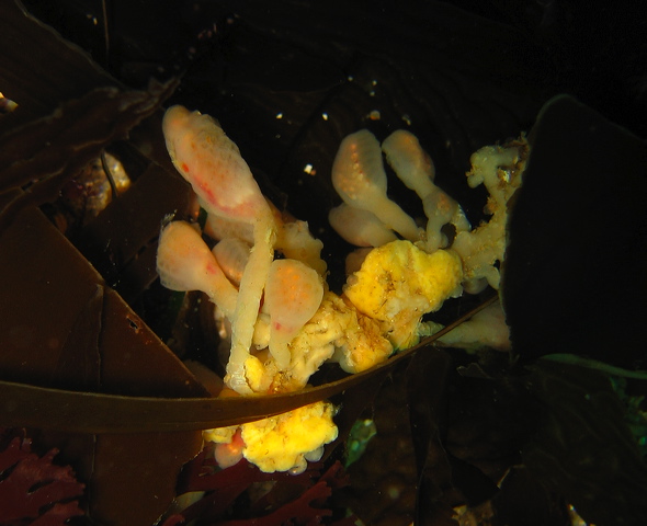
Polymastia pachymastia – Nipple Sponge
SP.?? - Sponge
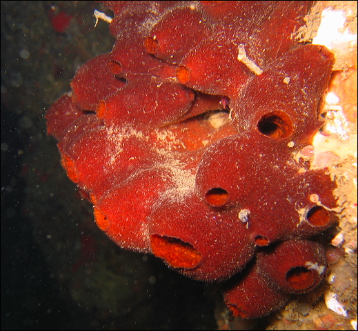
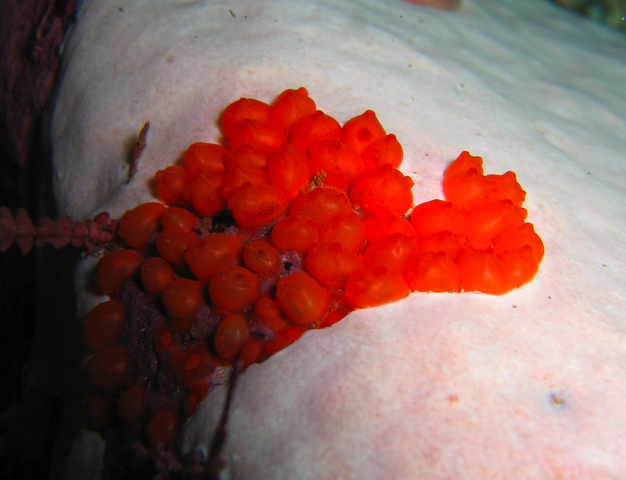
Acarnus erithacus – Red Volcano Sponge
Microcionia parthena – Red Upright Sponge 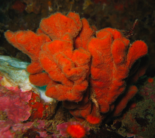
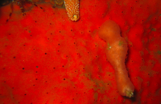
Red Encrusting Sponge
Hymenamphiastra cyanocrypta – Cobalt Sponge
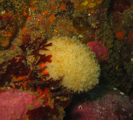
Halichondria sp. – White Urn Sponge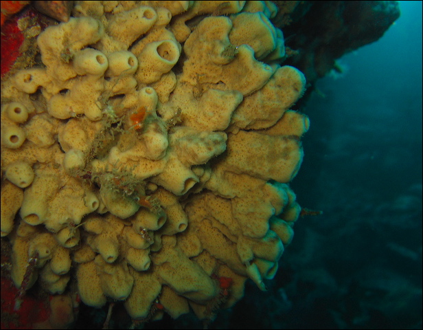
Brown Urn Sponge
Leucosolenia eleanor – Calcareous Sponge
Sponge sp.??
sp.?? - White Finger Sponge
The phylum Cnidaria is composed of seemingly quite unrelated groups of animals: anemones, hydrocorals, jellyfish. Some look like flowers attached to the surface, others are planktonic, freely floating in the water column.
Except for sponges, cnidarians have the simplest body structure: cells are organized only at the tissue level, there is no centralized nervous system, no head, no circulatory or sensory organs. Body parts are repeated around a central axis in radially symmetric fashion. Instead of front and back, oral (where the mouth is) and aboral (opposite to mouth) surfaces are distinguished. The centrally located mouth, surrounded by tentacles to capture prey, is the single opening to the gut (an incomplete gut lacking the anus) where food is digested. There are two body forms, a mirror image of each other: the sac like polyp and bell shaped medusa. In the polyps the oral surface is on top, tentacles oriented upwards, while in the medusa the tentacles trailing below. Some cnidarian groups have both forms, others are reduced to just one. With the invention of muscle and nerve tissues cnidarians are able to move and actively hunt. Because of their radial symmetry, however, movement is not directional, as they face their environment from all different sides. Cnidarians lack a hard skeleton, so for structural stability and rigidity they rely on a hydrostatic skeleton. The gelatinous middle layer, called the mesoglea, is composed mainly of water. As they lack respiratory and circulatory organs, gas exchange is done via diffusion through the body surface.
A defense common to all cnidarians is the nematocyst, a stinging structure produced by a venomous cell, the nematocyte are generally concentrated around the tentacles. Its function is to catch prey as well as to protect the organism from predators and fight off competition. In addition to delivering toxin, the nematocyst is occasionally used to inject digestive enzymes, as well as to capture debris to cover the animal. The nematocyst capsule contains a coiled hollow thread usually loaded with toxin that is ejected by water pressure as a response to a physical or chemical stimulus. Although all cnidarians have nematocysts, not all can penetrate human skin and their toxin potency varies. For example, moon jellies or many large anemones can be touched without feeling pain, but the box jelly is potentially lethal. Despite such chemical warfare several invertebrates and fish are predators of cnidarians.
- Hydrozoa
- Scyphozoa
- Anthozoa
- Staurozoa
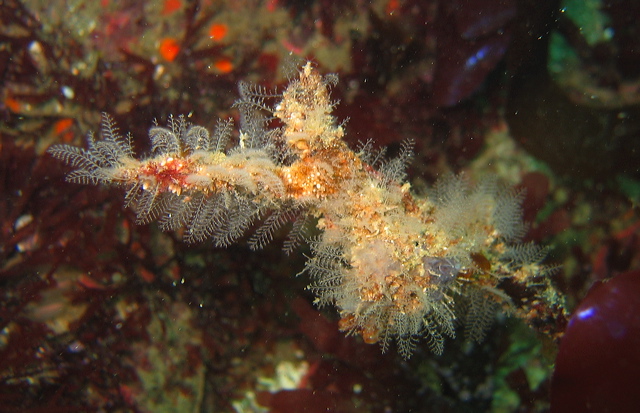
Hydrozoa
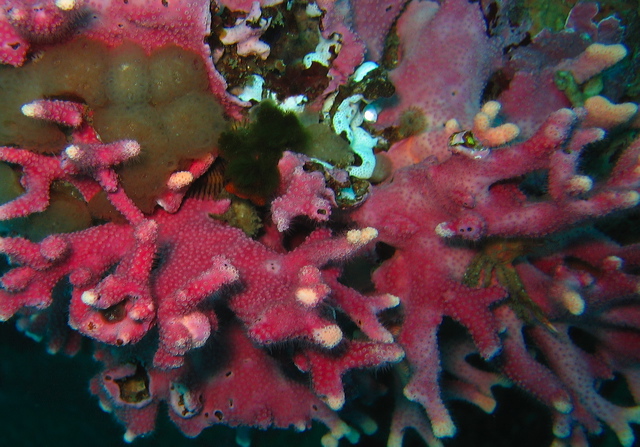
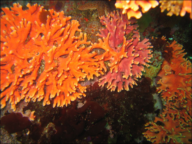
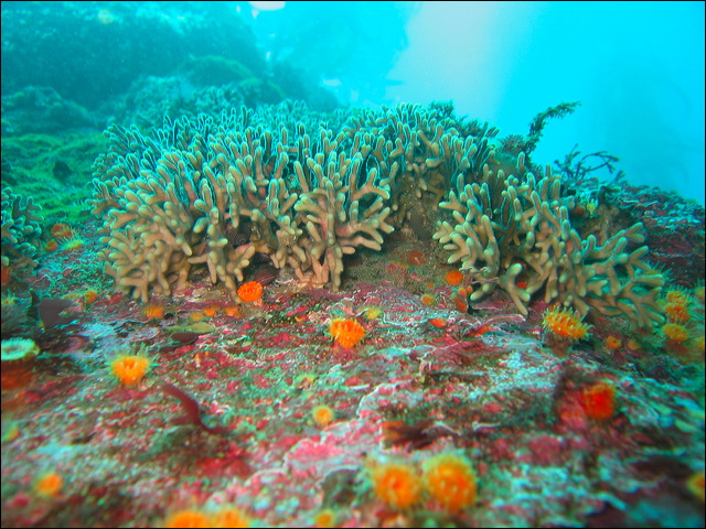
Most hydrozoa alternate between sexual planktonic medusa and an benthic polyp form, although some species may lack one or the other form. In the hydroid the polyp form is dominant. They are usually colonial, individuals are specialized for feeding, defense, and reproduction. The most visible hydroid in our waters is the California hydrocoral, both the encrusting and the upright form come in many shades of pink and purple. Hydrocorals secrete a calcium carbonate exoskeleton, hence the resemblance to reef building tropical corals. They are relatives of the tropical fire coral but with much less toxicity.
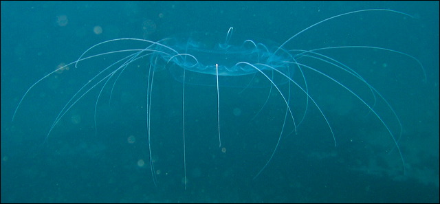
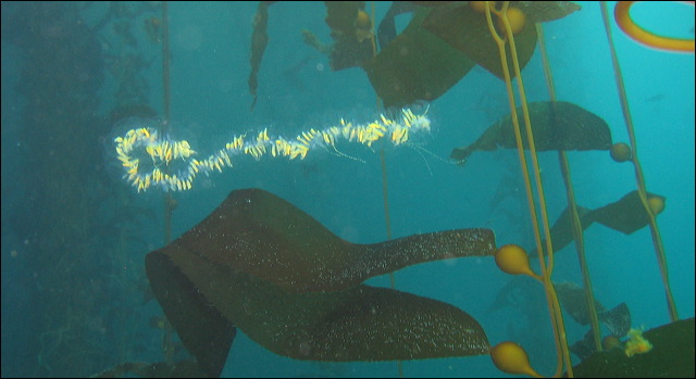
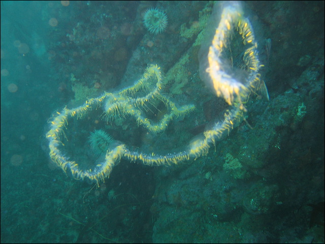
Hydromedusae live mainly in medusa form, somewhat similar to jellyfish. They are usually smaller, more transparent than jellyfish and have delicate tentacles. The characteristic thin circular flap within the margin of the bell, called a velum, distinguishes them form true jellyfish. Siphonophores are mobile hydrozoans that float in the water column, occasionally drifting close to shore. The most remarkable siphonophore, Praya, lives in the Monterey Canyon at great depth and can grow to an incredible length of 50 meters. A lucky diver may encounter a small fraction of it in shallow waters.
Aglaophenia struthionides – Ostrich Plume Hydroid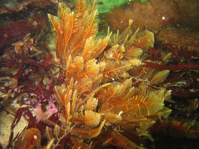
Hydractinia milleri – Hedgehog hydroid
Plumularia sp.
Stylaster californicus - California Hydrocoral
Annatiara affinis
Solmissus sp.
Scrippsia pacifica
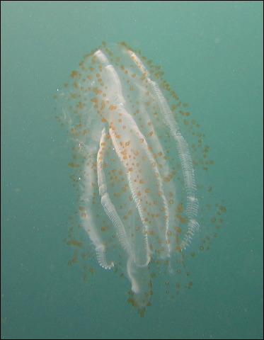
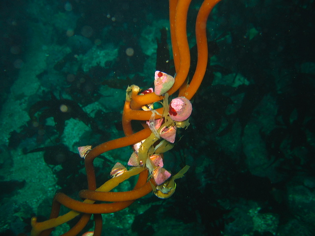
Siphonophore
Piece of a Siphonophore
Praya sp. - Pieces of a Siphonophore
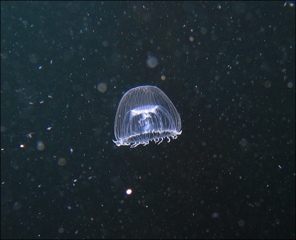
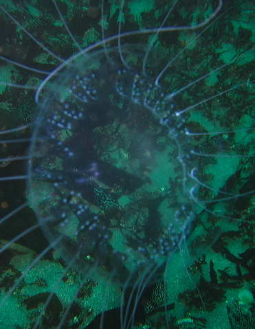
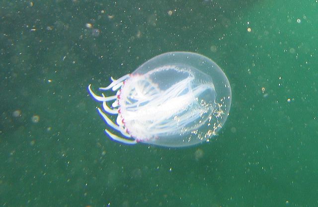
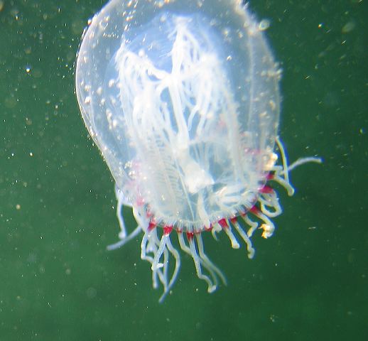
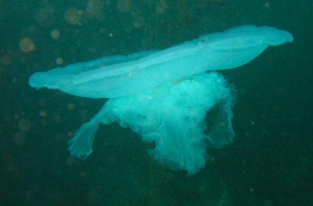
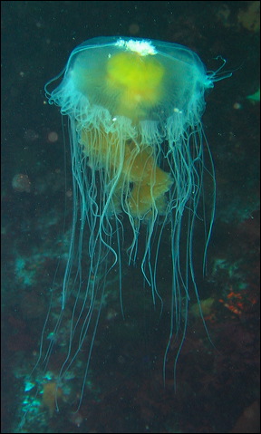
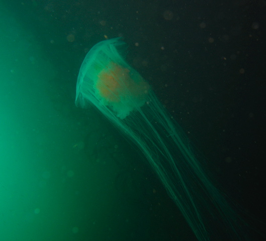
Scyphozoa – True jellyfish
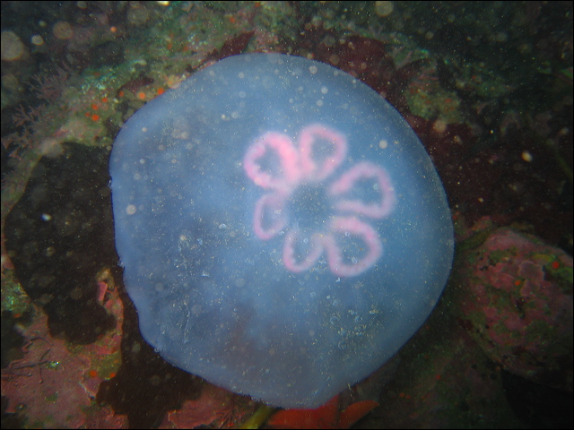
The medusa is the dominant stage in the life cycle of the jellyfish, but there is much more than what meets the eye. The life cycle of the moon jelly, typical to many other jellyfish, is an interesting story. The four horseshoe shaped pink or purple rings on the top of the white body are the reproductive structures, the starting point in sexual reproduction. Sexes are separate. The broadcast sperm is ingested by the female, fertilization takes place internally, and the female broods the planula larva. After a brief planktonic life the larva settles on a surface and metamorphoses into a polyp. The polyp reproduces asexually either producing new polyps by budding or new medusas by strobilation. In the latter process the polyp subdivides into a set of discs, which pop off at the end and become tiny juvenile medusas, called ephyrae, completing the cycle. While the medusa lives only for one season, the polyp may live more than a year, some even reach 20 years, and survive several strobilations. Such diverse life stages and reproduction strategies help the animal survive even in harsh conditions.
More than 95% of the body weight is water. Jellyfish are the favorite diet of the mola mola and some turtle species, even large anemonies play tug-of-war over a moon jelly. Though jellyfish lack true sensory organs, they have sensory centers, called rhopalia, connected to their nerve net located at the edge of the bell. At these spots they can sense light and dark with their non image forming eye spots, as well as perceive gravity to help in orientation. Moon jellies fly not only in our waters but also in orbit on board the space shuttle Columbia as subjects of a study on the effect of weightlessness on the development of organisms.
Aurelia aurita - Moon Jellyfish
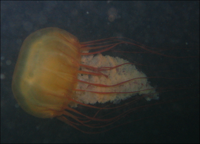
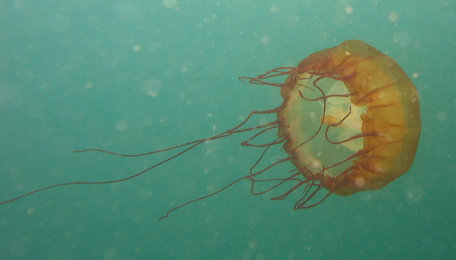
Chrysaora fuscesens - Brown Sea Nettle
Lots of tentacles
Bleached
No tentacles
Chrysaora colorata – Purple Jelly
Phacellophora camtschatica – Egg Yolk Jelly
Cyanea capillata – Lion's Mane Jelly
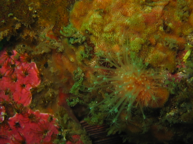
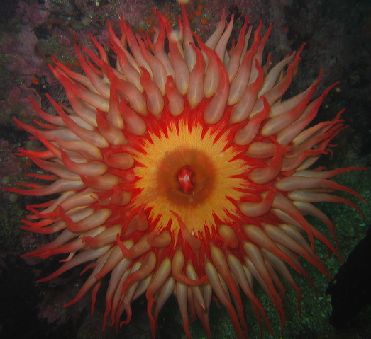
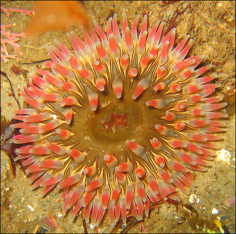
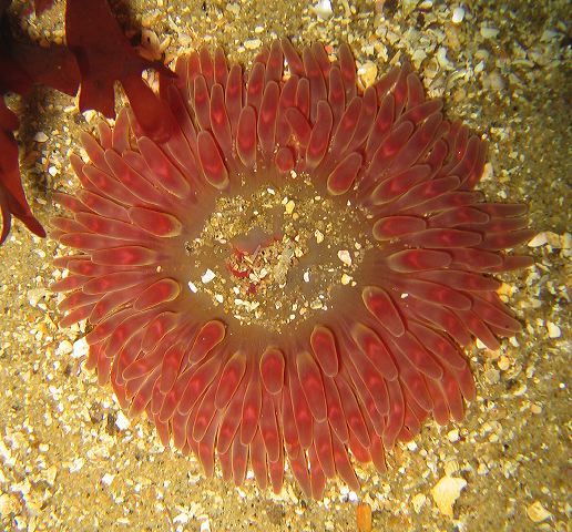
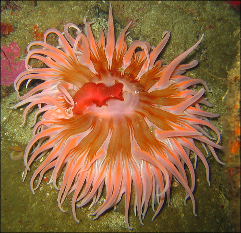
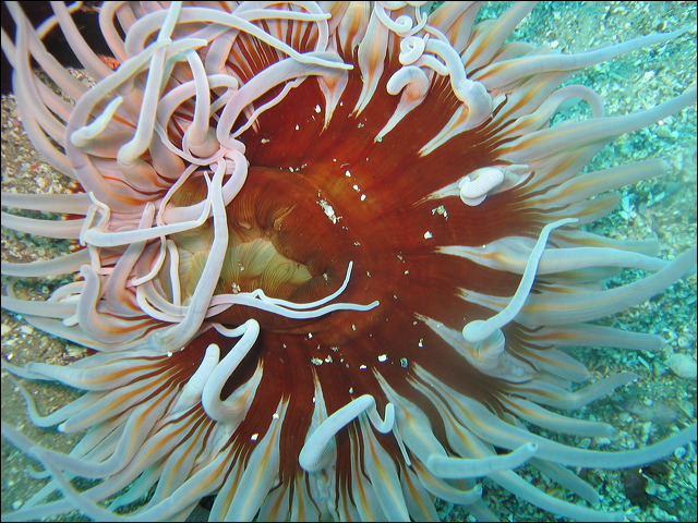
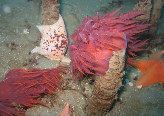
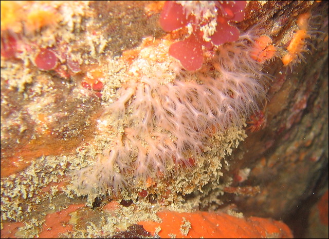
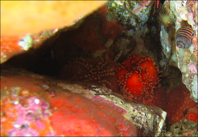
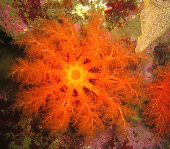
Anthozoa – Anemones, corals, sea pens
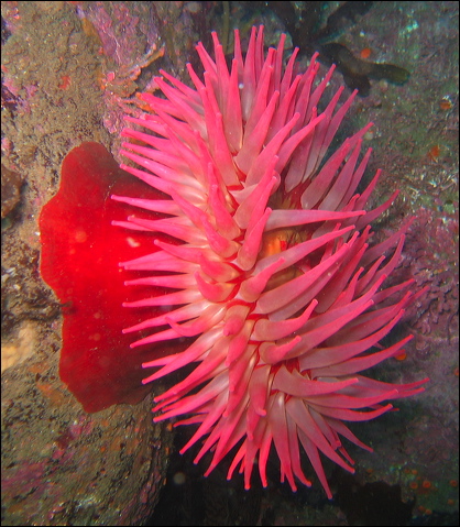
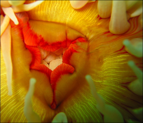
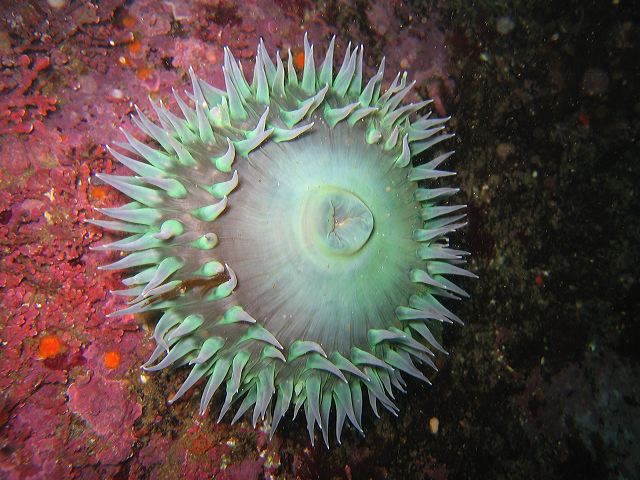
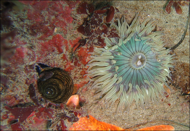
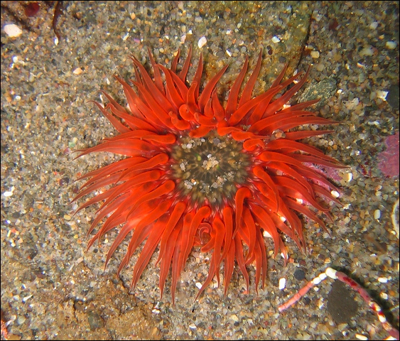
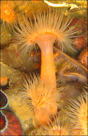
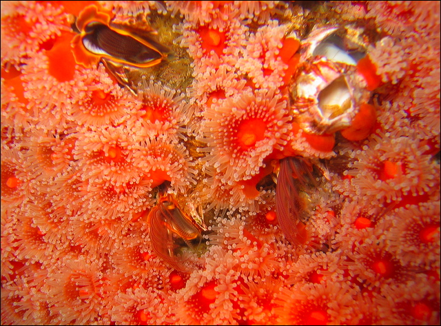
Anemones, named after a terrestrial flower, are indeed flower like creatures. True anemones live as large solitary individuals, but some species from related groups form a colorful colonial tapestry on the rocks. They are attached by the pedal disc and at the top of the cylindrical body is the oral disc with the mouth and stinging tentacles. They are all carnivorous catching live prey and paralyzing them with their nematocyst laden tentacles. Their diet consists of shrimps, crabs, chitons, snails, small fish, jellyfish, urchins. Unlike other Cnidarians, anemones lack the medusa stage, their life cycle has only polyps that can reproduce both sexually and asexually. Sexes are separate, some species broadcast sperms and eggs from the mouth others keep the fertilized eggs and develop them in brooding pouches. Asexual reproduction is done either by binary fission, i.e. splitting in half (e.g. Metridium) or just breaking off small pieces from the pedal disc, called pedal laceration. Anemones are mostly sessile, but are capable of creeping around on the basal disc, some can even completely dislodge and swim away if attacked.
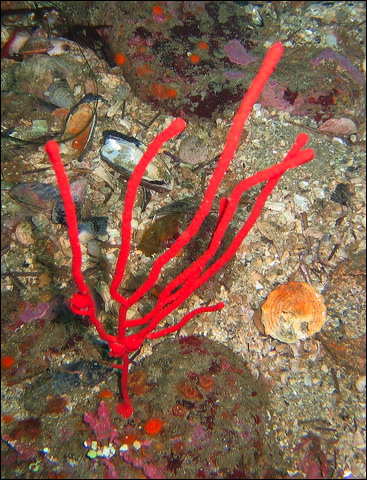
Anthozoans are divided into two subclasses: octocorals and hexacorals. Octocorals are colonial, the polyp has eight branched tentacles. This group contains soft corals, gorgonians and sea pens. Anemones and true corals with calcareous skeleton are hexacorals. With corals most of us associate spectacular reefs and large colonies. Corals in our waters are not reef builders and are rarely colonial. The only species that resembles tropical corals, the hydrocoral is in the Hydrozoa class.
Hexacorals
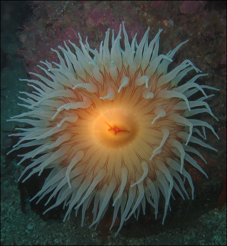
Urticina lofotensis – White Spotted Rose Anemone
Closed
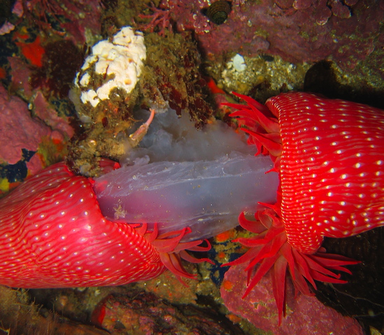
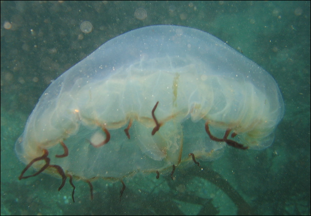
Eating a Moon Jelly
Urticina piscivora – Fish Eating Anemone
Urticina coriacea – Stubby Rose Anemone
Urticina columbiana – Sand Rose Anemone
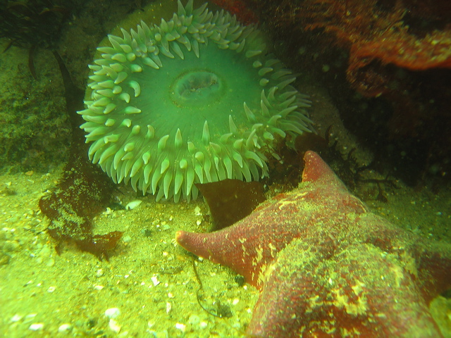
Anthopleura xanthogrammica – Giant Green Anemone
Anthopleura sola – Green Anemone
Anthopleura artemisia – Moonglow Anemone
Anthopleura elegantissima - Aggregating Anemone
Epiactis prolifera
Metridium senile - White Anemone
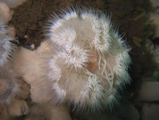
Metridium farcimen – Plumed Anemone
Cannery Point
Granite Point
Budding
Closed
Epizoanthus scotinus – Yellow Zoanthid

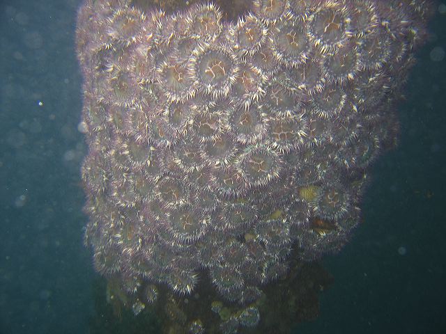
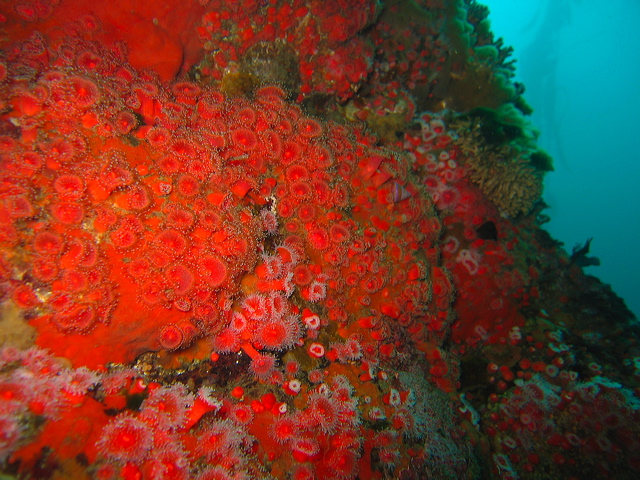
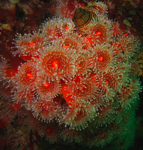
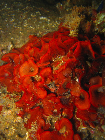
Corynactis californica – Club-tipped or Strawberry Anemone
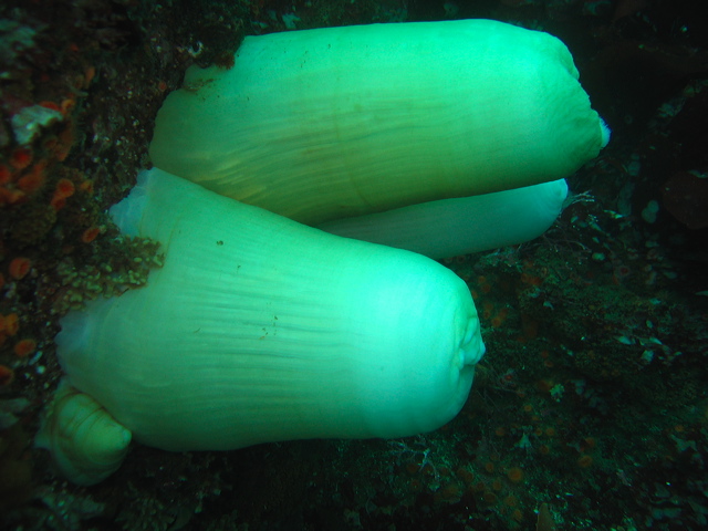
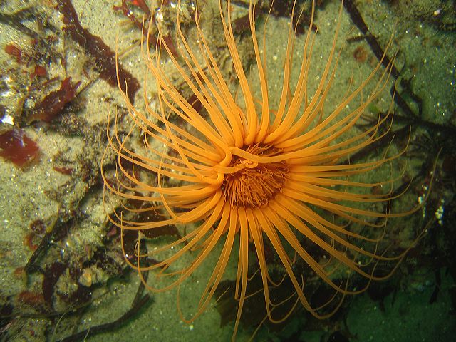
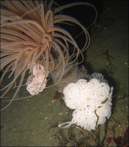
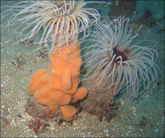
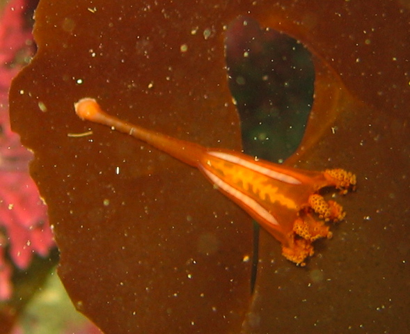
Pachycerianthus fimbriatus – Tube-dwelling Anemone
With Dendronotus Eggs
Tunicate Covering
With Dendronotus Eating
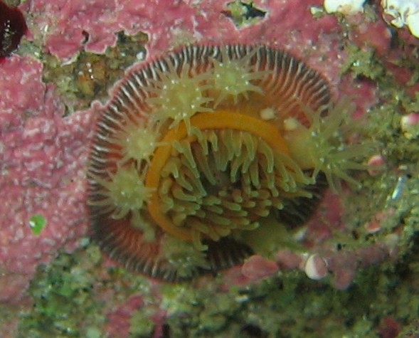
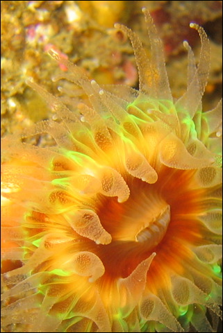
Paracyathus stearnsii – Brown Cup Coral
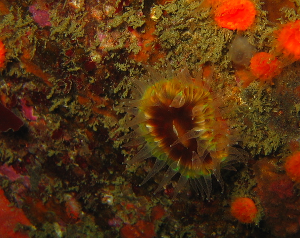
Balanophyllia elegans – Orange Cup Coral
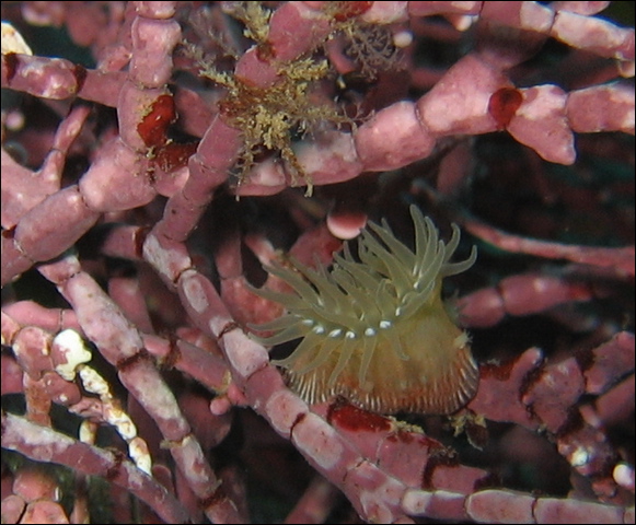
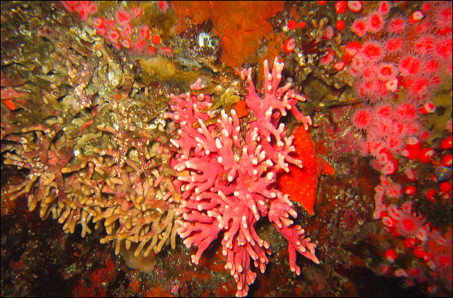
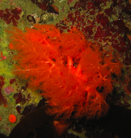
Octocorals
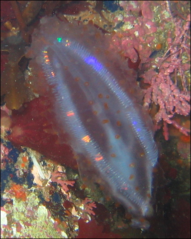
Stylatula elongata – White Sea Pen
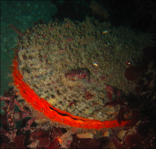
Ptilosarcus gurneyi - Orange Sea Pen
Clavularia sp. – Octocoral
Lophogorgia chilensis – Red Gorgonian
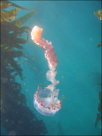
Manania handi – Stalked Jelly
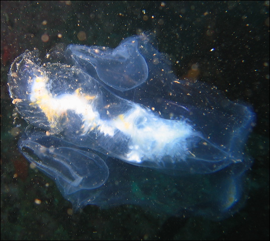
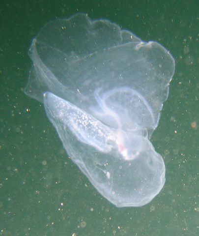
Comb jellies, also called sea gooseberries or sea walnuts, form the small phylum Ctenophora (meaning “comb-bearing”) containing only about 150 species, they are all marine. There may be only 150 species, but they are the most abundant among all the gelatinous animals, they form a major portion of the planktonic biomass. The radially symmetrical, transparent, gelatinous body resembles the true jellies (Scyphozoa), but there are some significant differences between these two planktonic groups. On average comb jellies are small (few centimeters), spherical or ovoid in shape. They lack stinging cells (nematocytes), instead they have sticky cells, called colloblasts. These carnivorous predators capture their food with two long, retractable tentacles laden with colloblasts. In contrast to the pulsating jellyfish, comb jellies propel themselves with their eight rows of “combs” (ctenes), that are aggregations of large cilia. These vigorously beating combs reflect light, filling the eight rows with rainbow colors. The comb jelly’s nervous and digestive systems are more developed than in true jellies, but gas exchange and excretion happens through simple diffusion over the entire body surface. Similar to true jellies the statocysts, found at the aboral top end, control balance and orientation. The life cycle of comb jellies is simple, without medusa – polyp alternation and without attached sessile life stage. Most species are hermaphrodites, even capable of self-fertilization. Sexual reproduction is the norm, fertilization takes place in the water column using broadcast fertilisation. Comb jellies are able to regenerate any lost part, even an entire half of the body.
Beroe sp. - Comb Jelly
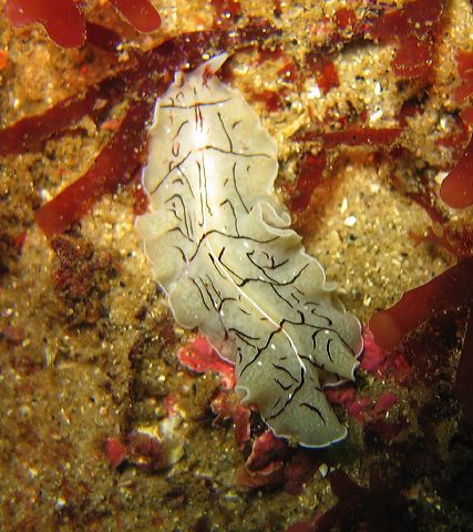
The word ‘worm’ is for most of us associated with ugly, slimy creatures. But worms in the marine environment certainly do not fit this stereotype. There are several phyla of worms: flatworms, ribbon worms, roundworms, peanut worms, segmented worms. In terms of evolution the most exciting perhaps is the phylum Platyhelminthes, the flatworms. Their body plan is a major developmental step from Cnidarians as they display bilateral symmetry, a head with a simple brain, and true organs. However, it is quite rare to encounter a flatworm because they are well hidden under rocks usually in shallow waters.
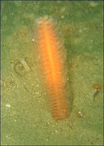
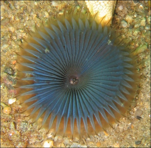
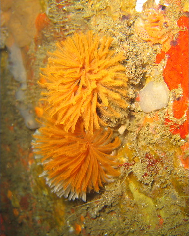
A group of segmented worms (Annelida), the Polychaete worms, are the ones we encounter most frequently, but they are cryptic, so you have to look for them on kelp holdfasts, on sandy bottoms, in between the folds of sponges and tunicates. Their bilaterally symmetrical body consists of segments, as a series of similar parts. Each segment has a pair of extensions, the parapodia, with sharp bristles that are used for both locomotion and gas exchange. The parapodia, however are greatly reduced in the tube-dwelling worms. There is a head with a brain, eyes and other sensory organs. They have a complete gut and a closed circulatory system transporting nutrients as well as oxygen and carbon dioxide. Waste is removed by the excretory organs and gas exchange is accomplished with gills. These worms have quite a complex architecture.
Many polychaetes are filter feeders, others are deposit feeders or active predators. As several species in our waters are tube dwellers, their movement is restricted and what we see are the tentacles covered with cilia that stretch out to capture and transport small invertebrates. In addition to feeding, these tentacles are also used in gas exchange. In some species, e.g. in christmas tree or feather duster worms, these tentacles are arranged in beautiful complex shapes. Mucus is used not only in helping to catch food but also to cement the tubes built from seaweed, mud, sand and shell fragments. Some species are able to not only retract their tentacle but can also close their tube with an operculum.
Polychaetes have separate sexes, fertilization occurs in the water column after broadcasting eggs and sperms. The trocophore larvae of the polychaetes are similar to the larvae of molluscs. They also have various degrees of regenerative capabilities, most can regrow posterior body segments, some even anterior segments including the head. The most impressive is a Dodecaceria species that can fragment itself into individual segments and regenerate an individual from each segment.
Phylum Platyhelminthes – Class Turbellaria – Order Polycladida
Pseudoceros luteus
Eurylepta californica
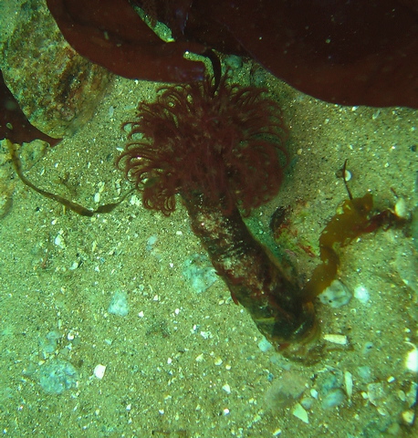
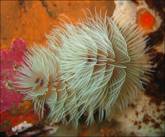
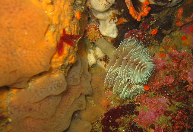
Phylum Annelida - Class Polychaeta - Order Sabellida - Family Sabellidae
Myxicola infundibulum – Sabellid Worm
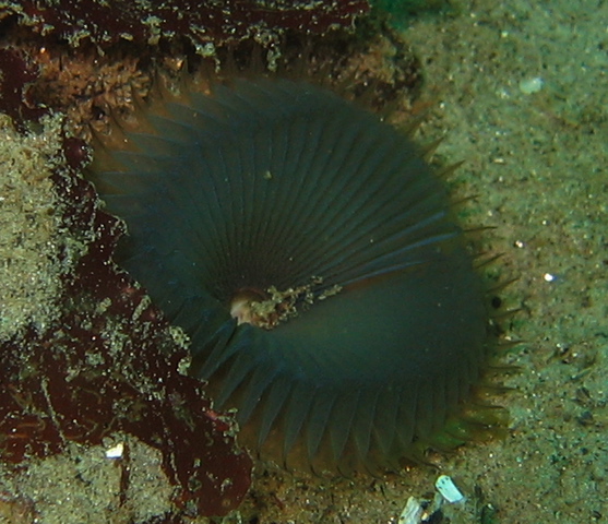
Eudistylia polymorpha – Feather Duster Worm
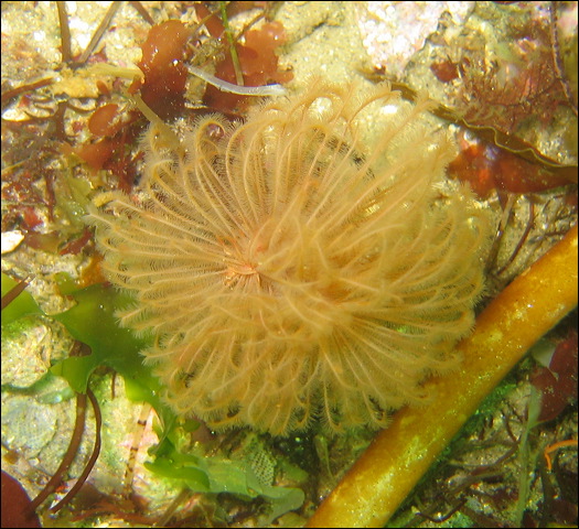
Eudistylia vancouveri - Northern Feather Duster Worm
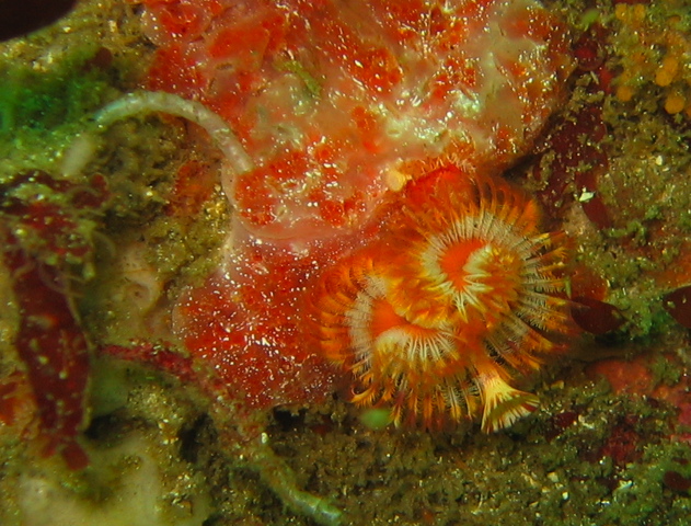
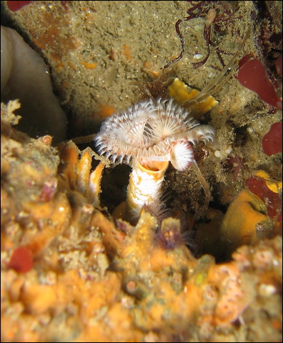
Phylum Annelida - Class Polychaeta - Order Sabellida - Family Serpulidae
Serpula vermicularis – Serpulid Worm
SP.?? – Worm
Phylum Annelida - Class Polychaeta - Order Cirratulida - Family Cirratulidae
Dodecaceria fewkesi - Colonial Tube Worm
Dodecaceria concharum – Coralline tube worm
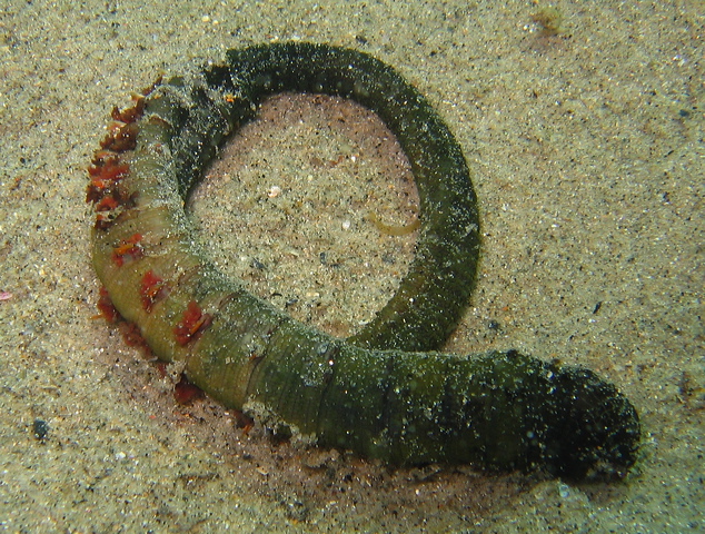
Arenicola marina - common Lugworm (normally buried in the sand)
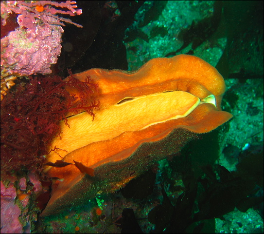
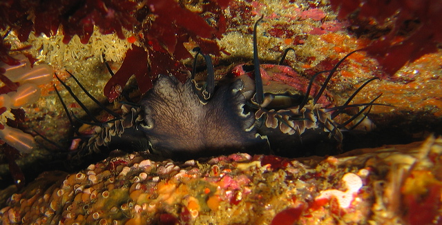
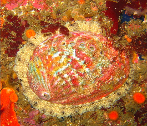
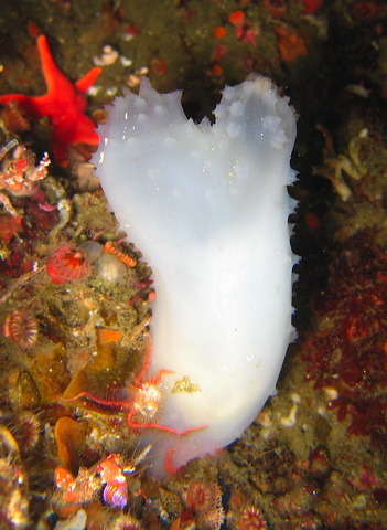
The phylum Mollusca contains the highest number of species in the ocean among all animal phyla. This group contains the most familiar invertebrates: snails, clams, oysters, scallops, squids, octopuses. They are harvested, even farmed for human consumption and their shells are collector items. Nudibranchs, the favorite subjects of many underwater photographers, are also members of this group. The size range is huge, from tiny bivalves to the giant octopus and squid. Most species we encounter under water belong to four classes: gastropods (snails, slugs, abalone, limpets), bivalves (clams, mussels, oysters, scallops), cephalopods (squid, octopus), and chitons.
The soft mollusc body has three general regions: head, foot, and visceral mass. The head is equipped with sensory structures: eyes, statocyst, tentacles. The large muscular foot is a key structure for locomotion. The body is covered by a thick sheet, called the mantle, it forms a cavity, the mantle cavity, that houses the gills. In most species the mantel secretes a shell, or shell plates. A characteristic structure in most molluscs is the radula, a tongue like belt with chitinous teeth used in scraping food from surfaces. The number and shape of these teeth are important keys in grouping of molluscs. Some species incorporate a large amount of iron in the radula. The open circulatory system includes a heart that is composed of separate ventricle and atria. A complete gut performs digestion, a large and complex kidney is responsible for excretion, while gas exchange is done through a pair of gills. Their nervous system is well developed and centralized in a brain. Most molluscs have separate sexes, but there are exceptions most notably the nudibranchs that are hermaphrodites. Adults develop from the characteristic trochophore larva, some form an additional larval stage, called veliger.
- Chitons
- Gastropods
- Bivalves
- Cephalodpods
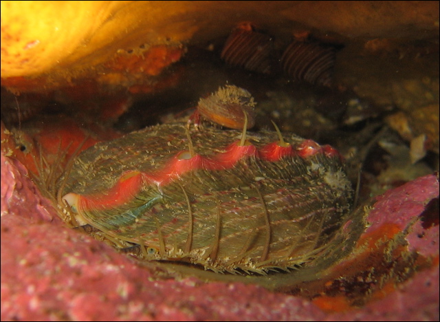
Chitons
Chitons form an exclusively marine class, its name, Polyplacophora, means “many plate bearer”. Their architecture is quite primitive compared to other molluscs. The flattened, elongated body has a ventral foot and eight dorsal shell plates that are embedded in a thickened region of the mantle, called the girdle. Sometimes the plates are partially or totally covered by the girdle. The butterfly shaped plates, often found washed up on beaches, give their body flexibility, so that they can roll into a ball when attacked. They have no tentacles, but can sense light with ocelli. Almost all chitons are herbivores. Chitons are often found in intertidal areas. The largest species is the gumboot chiton, commonly found in our waters, its eight plates are completely covered by a redish brown mantle.
Tonicella lineata – Lined Chiton
Cryptochiton stelleri – Gumboot Chiton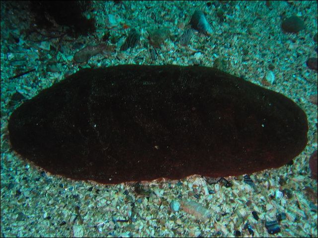
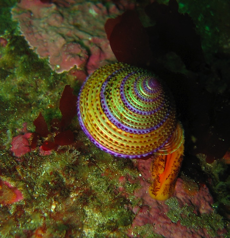
Gastropods
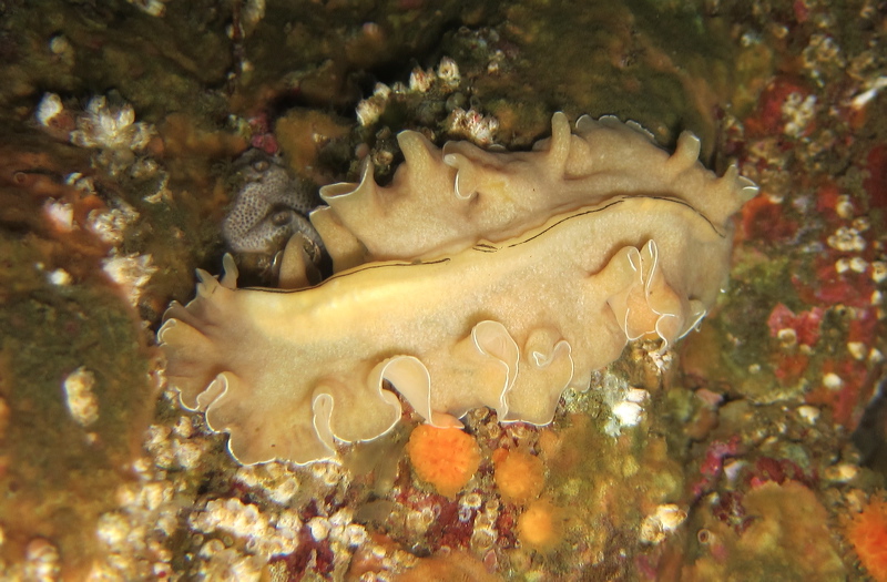
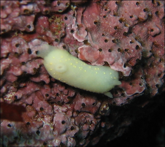
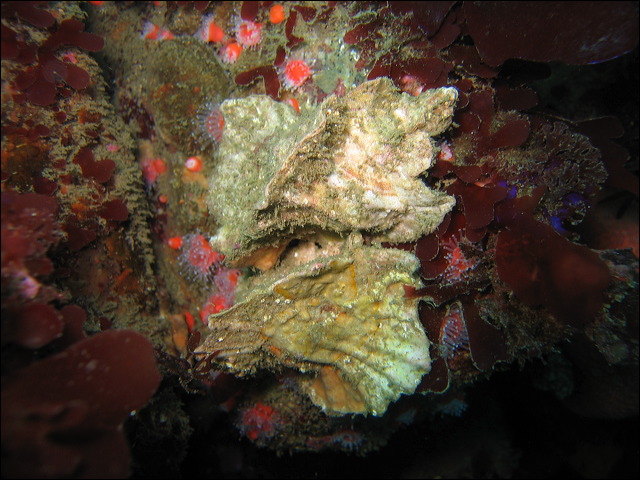
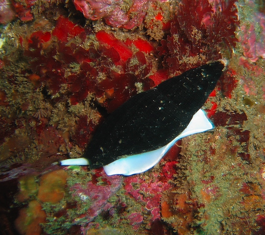
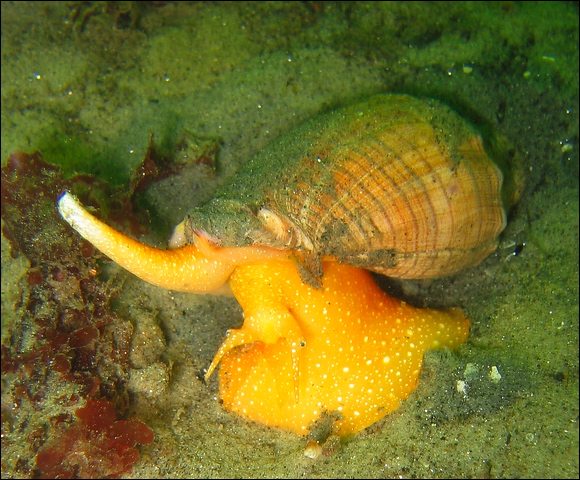
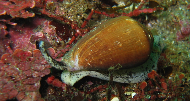
The Latin name of the class, Gastropoda, means “stomach footed”. These well known mollusc species, snails and slugs, are common in water (salt and fresh) as well as on land. Many animals in this group of molluscs under go torsion, a strange 180 degrees twist of the body above the foot, unique in the animal kingdom to gastropods. Torsion takes place during development in the veliger larval form. This results in an asymmetric structure, a coiled mass of organs enclosed in a shell above the foot, where the anus is located just above the head. There are several theories, but none explains fully the reason for this unusual developmental step. Several gastropods are herbivores, scraping algae from rocks, many, e.g. the cone shell, are carnivores attacking worms and bivalves, others, like whelks feed on detritus (dead animals) using their proboscis, a tubular sucking organ. In nudibranchs the shell is absent, hence the name “naked gills”. They prey on sponges, tunicates, bryozoans, hydroids. Without the protective shell they rely on noxious chemicals, advertised by their bright colors, and even retain nematocysts (stinging structures) from their prey cnidarians.
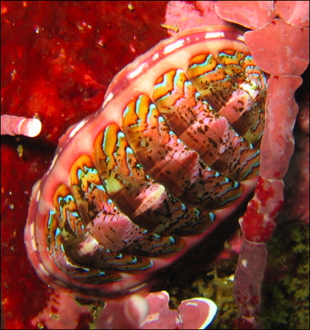
Haliotis rufescens - Red Abalone
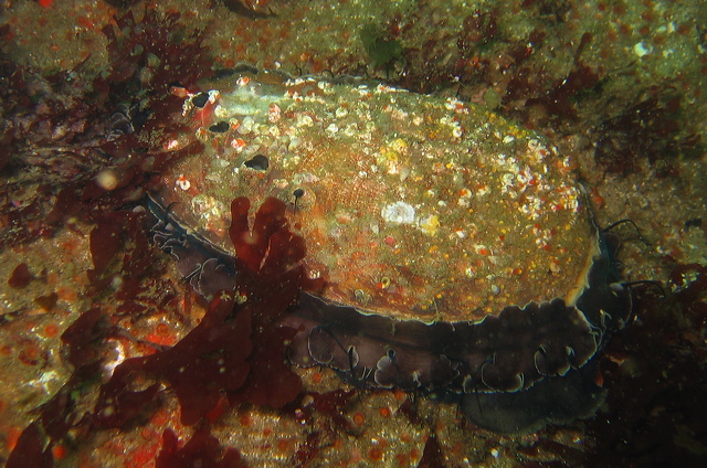
Haliotis walallensis – Flat Abalone
Young flat abalone
Diodora aspera – Rough Keyhole Limpet
Tegula montereyi
Megathura crenulata – Giant Keyhole Limpet
Calliostoma annulatum – Blue Ring Top Snail
Calliostoma ligatum – Blue Top Snail
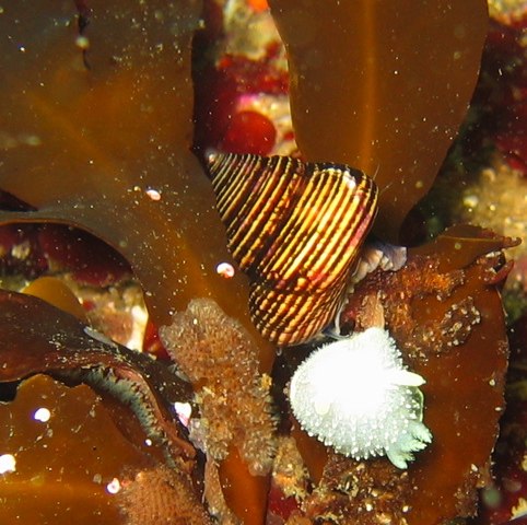
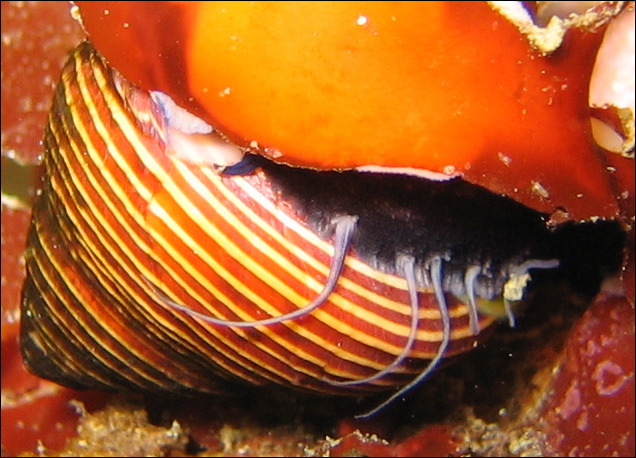
Lithopoma gibberosa - Red Turban Snail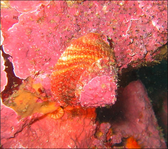
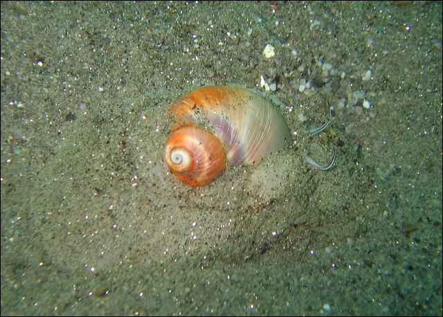
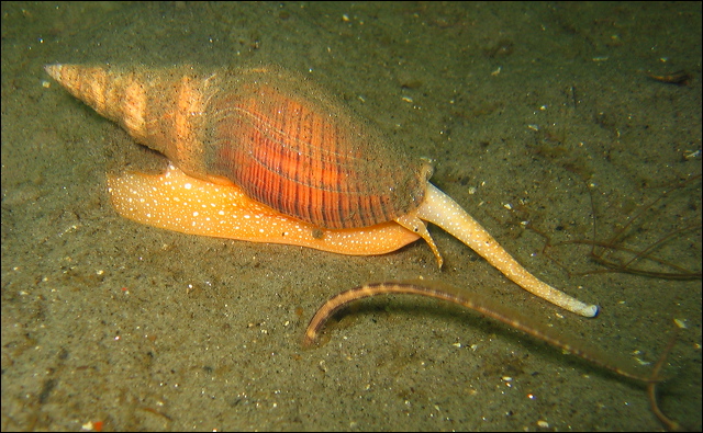
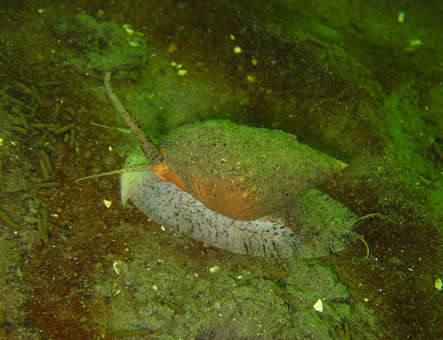
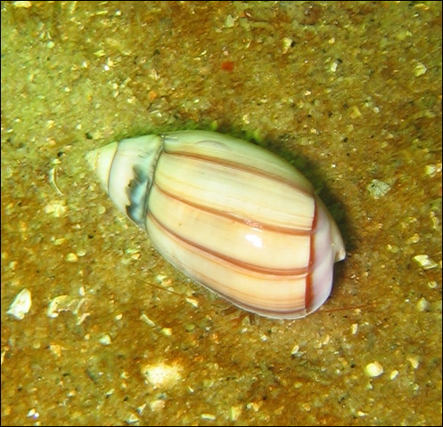
Euspira lewisii – Lewis’ Moon Snail
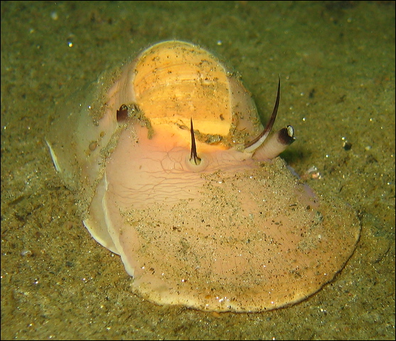
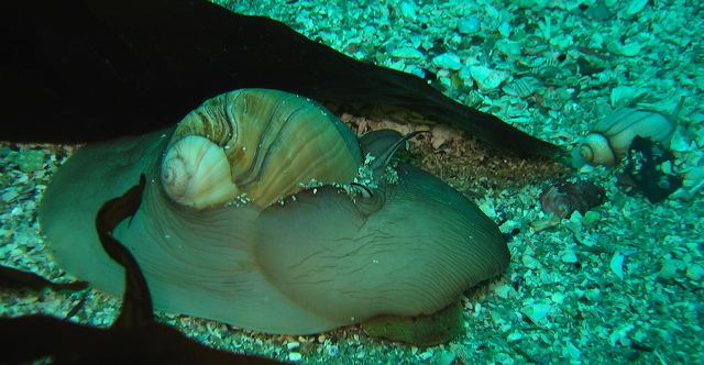
Lewis' Moon Snail eggs mixed with sand and then formed around shell.
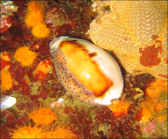
Cypraea spadicea – Chestnut Cowry
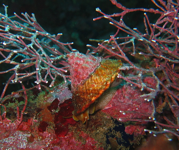
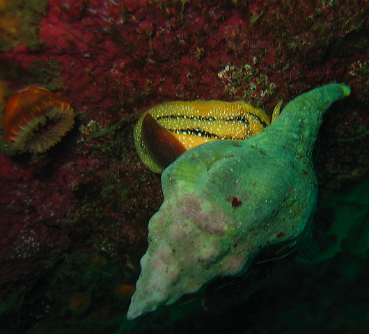
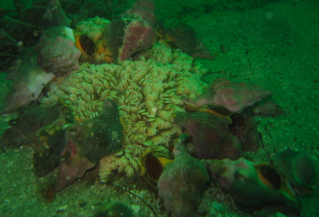
Kelletia kelletii – Kellet’s Whelk
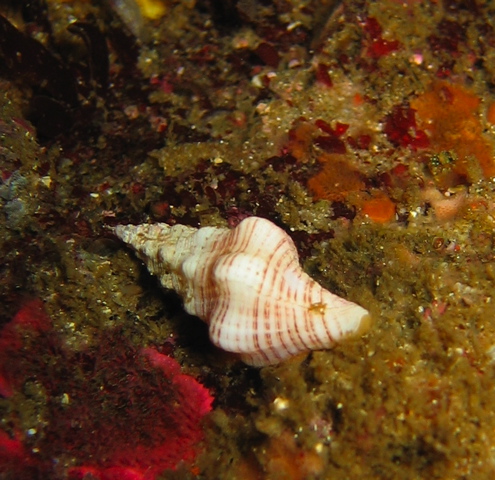
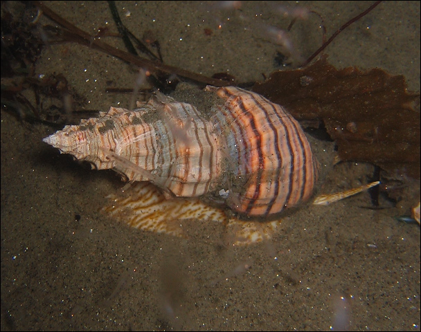
Ceratostoma foliatum – Leafy Hornmouth
Mitra idae – Ida’s Miter
Megasurcula stearnsiana
Conus californicus – California Cone Snail
Cancellaria cooperi - Coopers Nutmeg
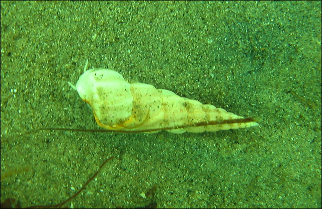
Nassarius fossatus - Channeled Nassa (or whelk)
Olivella biplicata - Purple Olive Snail
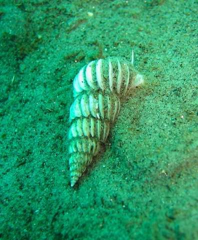
Opalia borealis - Boreal Wentletrap
Family: Cavoliniidae
Corolla Calceola (The specimen is incomplete)
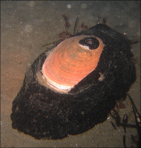
Bivalves
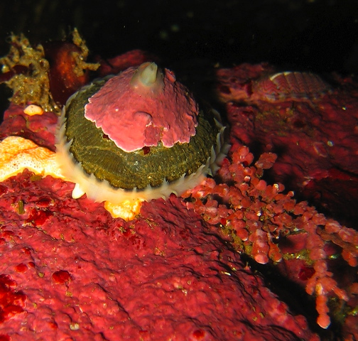
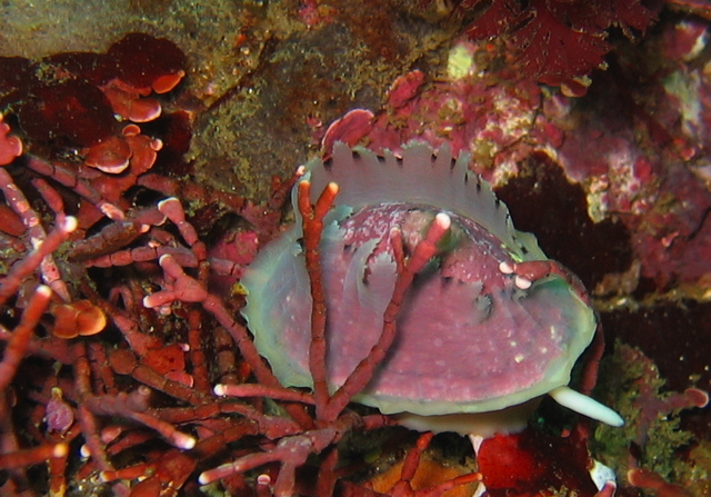
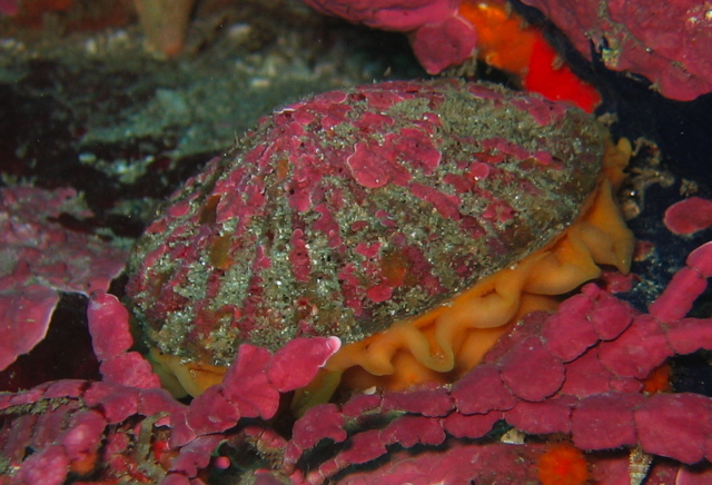
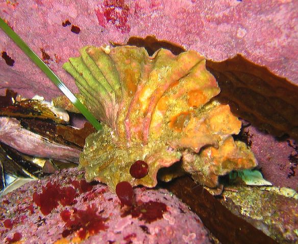
Bivalves (mussels, clams, oysters, scallops) have a laterally compressed body inside a pair of shells, called valves. The valves are hinged together by ligaments and a tooth like structure, and closed together by adductor muscles. Each valve has a dorsal hump close to the hinge, called the umbo, the oldest part of the shell from where concentric growth lines radiate. The shells are lined by the mantle, so the entire body lies in the mantle cavity. When a foreign object, like a sand grain lodges between the shell and the mantle, a pearl is formed as the animal coats the irritating particle. There is no head and no radula. Instead of image forming eyes they sense light with eye spots, called ocelli, often seen around the edge of an open scallop. Bivalves are filter feeders using their gills to catch food particles as well as for gas exchange. Most bivalves live attached to a surface or buried under the soft bottom. Oysters cement their shell to a rock, mussels attach themselves by byssal threads. Some scallops can swim by clapping their valves and ejecting water from the mantle cavity. Some clams live in the mud, using their shovel shaped foot to bury themselves and their siphons to draw in water.
Crassadoma gigantea – Rock Scallop
Juvenile recently settled.
Parapholas californica – Scaleside Piddock
Chaceia ovoidea – Wartneck Piddock
Panope generosa – Geoduck Clam
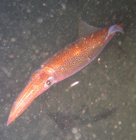
Cephalopods
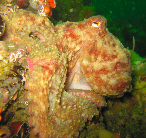
Cephalopods are the most developed, “intelligent” invertebrates, their nervous system is the most advanced of all invertebrates. In addition to squids and octopus, that can be seen in our waters, this class includes cuttlefish and the famous nautilus, the only cephalopod with an external shell. The giant squid is the largest living invertebrate exceeding 20 meters. In squid the shell is reduced to the internal pen, while it is absent in octopuses. Their body architecture is modified for an active predatory life style. All are efficient swimmers, they can propel themselves in any direction by forcing water through the siphon, which can be moved around. The foot has been modified into arms and tentacles equipped with suckers. The eyes are well developed and can form images. Indeed the squid posses one of the largest eyes for its size known in the animal kingdom. Unlike other molluscs, cephalopods have a closed circulatory system and a three part heart pumping blood separately to gills and body. One of the arms of the male is modified to transfer sperm packets into the mantle cavity of the female. For camouflage and communication cephalopods, including octopus, use dramatic color displays activating their chromatophors, pigments in the skin. Their beak, often venomous, is the only hard part in the soft body, that is why they can squeeze into small spaces. Releasing ink is a unique way to distract predators. Cephalopods live a very short life, even the giant octopus lives only 3- 4 years, they die shortly after mating.
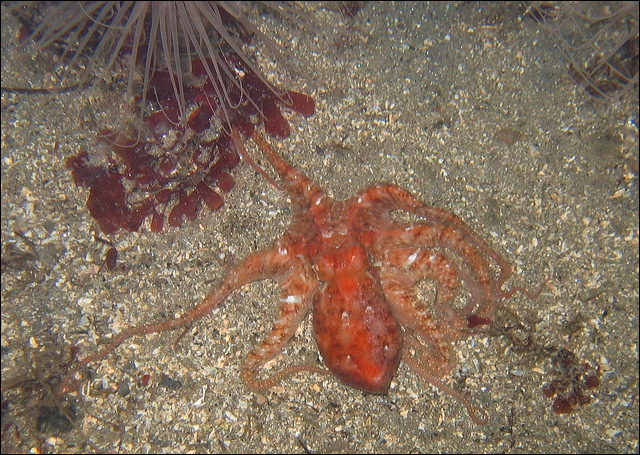
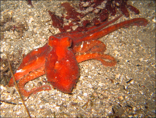
Octopus rubescens – Red Octopus
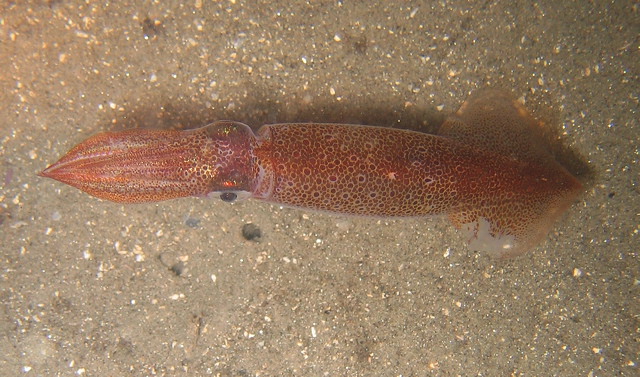
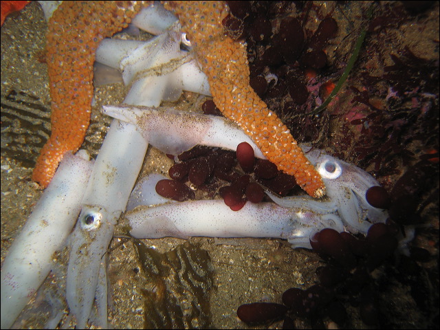
Loligo opalescens - Market Squid
Dead
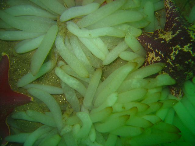
Squid Eggs
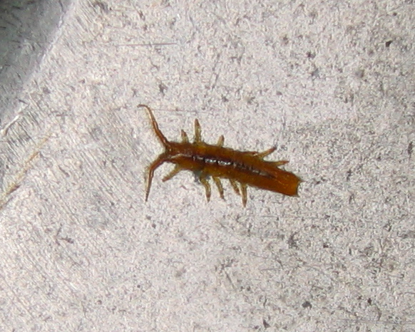
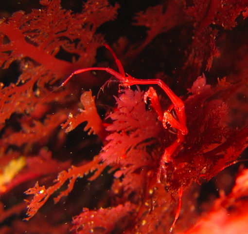
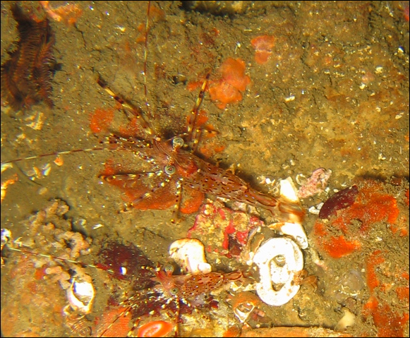
Arthropoda is the largest phylum of animals, comprising about 80 percent of all animal species. The Greek name, jointed feet, refers to one of the primary characteristics of the group. Insects, rare in the ocean, are the most abundant arthropods. Most marine arthropods, for example, shrimp, crab, lobsters and barnacles are crustaceans, members of the subphylum Crustacea.
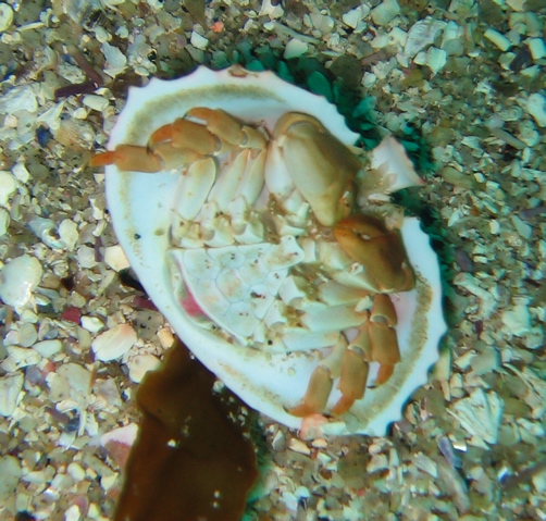
The crustacean body is bilaterally symmetrical and segmented, typically divided into a head, thorax, and abdomen. In true crabs the abdomen is tucked under the thorax, V shaped in male, U shaped in female. Each segment is equipped with jointed appendages used in locomotion, feeding, defense, or mating. The most visible feature is the non-living exoskeleton (external skeleton) that provides protection, support and flexibility. The part of the exoskeleton covering the head and the thorax is called the carapace. The exoskeleton is made up of chitin a form of carbohydrate polymer, it is a hard substance very similar in strength to fingernails (although fingernails are made of keratin). The exoskeleton is a limitation to the size of crustaceans they are never as large as some other invertebrates, for example cnidarians or molluscs. As the animal grows, and the shell becomes too small, it must be shed and replaced. This process is called molting or ecdysis. Before discarding the old shell a new soft shell is secreted by the underlying tissue. When the animal has molted the old shell it puffs up its body with water to ensure that the new shell hardens to a comfortably large size. It is possible to find numerous empty shells at most dive sites, they can just look like the original animal, perfect in every detail except the animal has left.
Small planktonic crustaceans are key components near the base of the food chain. Copepods provide the link between phytoplankton (floating organisms capable of photosynthesis) and the zooplankton. Some copepod species are the most abundant animals on earth, they are the biggest protein source in the ocean. The planktonic, shrimp-like krill, feeding on phytoplankton, are abundant in polar waters and provide food for many species of whales, penguins, fish. The biomass of the Antarctic krill is the largest of all marine animals. Amphipods are laterally flattened, small crustaceans. One benthic species is the main food source for Gray whales. Skeleton shrimp are a subgroup of amphipods, their slender transparent body can be seen attached to seaweed, hydroids, or bryozoans. The parasitic fish lice is an isopod, so is the small, flat, kelp-colored animal stuck to your dry suit as you exit from the water thick with kelp.
The order Decapoda includes the largest crustaceans. This group contains commercially important invertebrates like shrimp, prawn, lobster and crab. The name refers to the five pairs (total of ten) of walking legs. The first pair, the claws, called chelae, are usually larger and tend to be used in feeding and defense. The small appendages attached to the abdomen, called pleopods, are used for swimming and brooding eggs, while the three pairs close to the mouth are the maxillipeds that sort food particles and pass them to the mouth. Two pairs of antennae on the head help sensing the environment. In addition to compound eyes, often elevated on stalks, there are several light sensing eye spots, called ocelli. Decapods have an open circulatory system with a dorsal heart, and a complete gut with digestive glands. Gas exchange is performed via gills located under the carapace. The ventral nervous system is centralized in a brain. Usually sexes are separate, the male uses specialized appendage to transfer sperm into the female. Mating usually takes place right after the female has molted and eggs are often brooded on the pleopods. The planktonic larvae undergo several molts, each adding a new appendage.
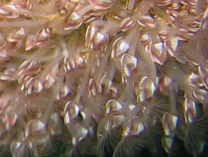
Barnacles live attached to surfaces, not only rocks but also other animals, e.g. whales. Some species are the most aggressive fouling organisms and they can be found on docks and boats. In the intertidal zone barnacles compete for space with snails and limpets. Barnacles are mistakenly thought of as mollusks as they live inside a calcareous shell. They are sessile (non moving), glued head down to the surface, only their feathery legs are sticking out from the opening of the plates filtering the sea water. All barnacles are hermaphroditic.
Idotea sp. - Isopod
Pollicipes polymerus – Goose Barnacle
Balanus nubilus – Giant Acorn Barnacle

Megabalanus californicus – Barnacle
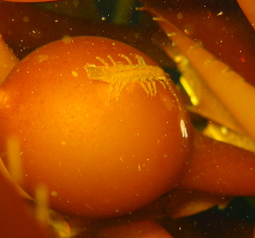
Mysid
Caprella sp. - Skelton Shrimp
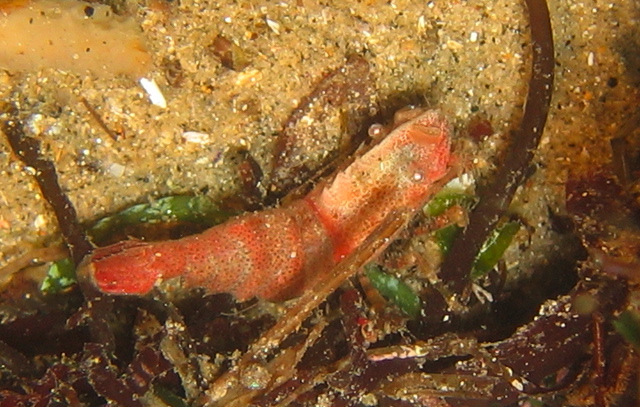
Crangon sp.
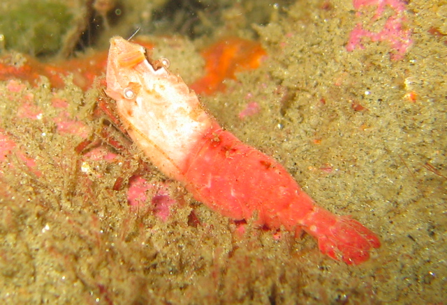
Pandalus gurneyi - Dock Shrimp
Pandalus platycerus – Spot Prawn
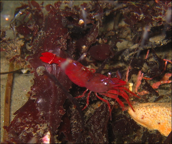
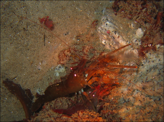
Heptacarpus palpator
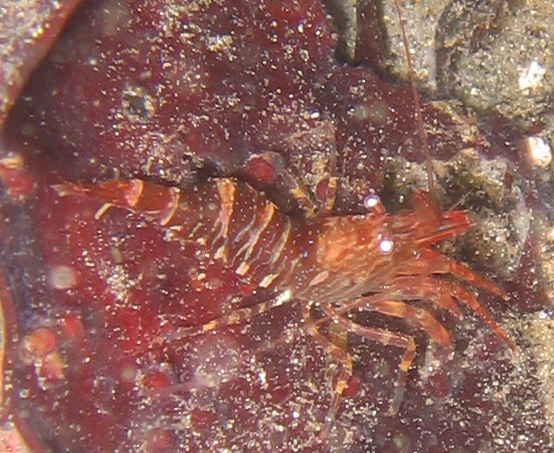
Heptacarpus stylus - Stiletto Shrimp
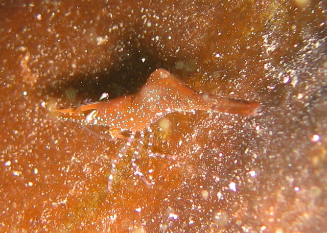
Spirontocaris prionota
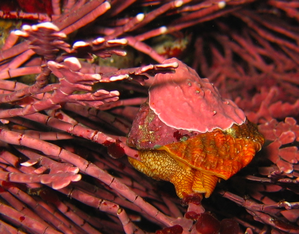
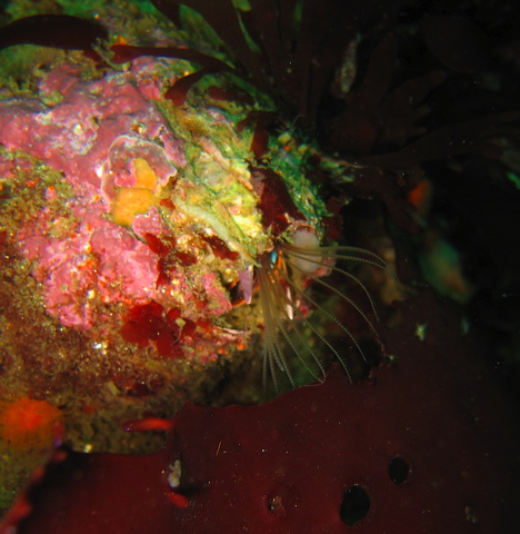
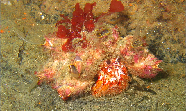
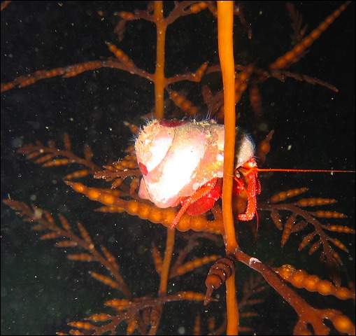
Pagurus hemphilli – Hermit Crab
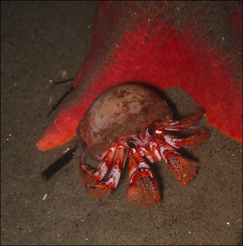
Pagurus armatus – Hermit Crab
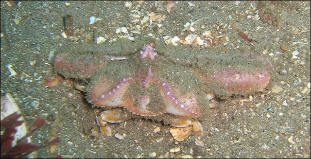
Cryptolithodes sitchensis - Umbrella Crab
Phyllolithodes papillosus - Lithoid Crab
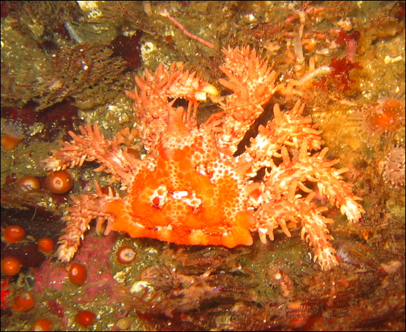
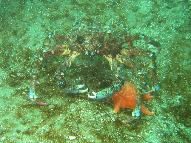
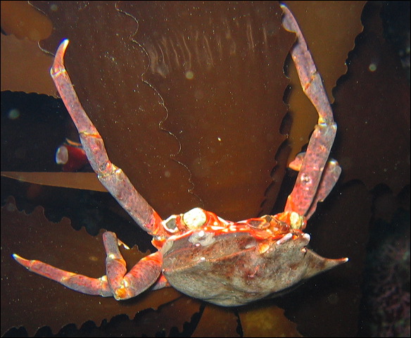
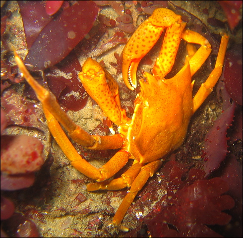
Loxorhynchus crispatus - Masking Crab
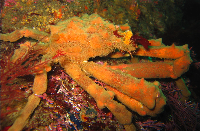
Loxorhynchus grandis – Sheep Crab
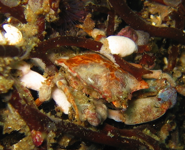
Mimulus foliatus - Mimicking Crab
Pugettia richii – Kelp Crab
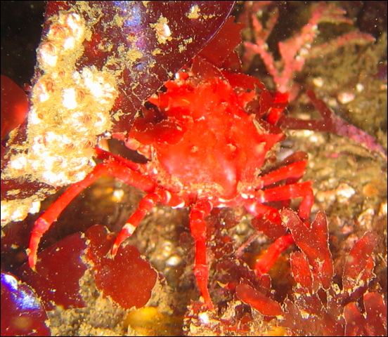
Pugettia producta – Kelp Crab
Heterocrypta occidentalis – Elbow Crab
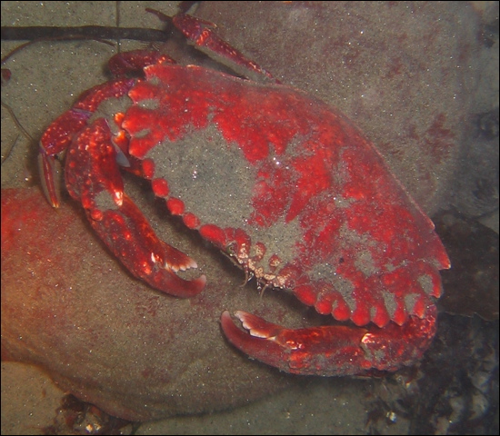
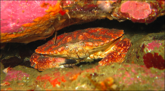
Cancer antennarius - Brown Rock Crab
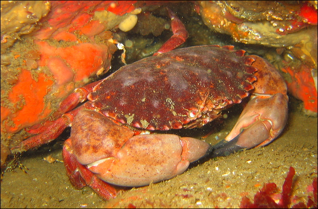
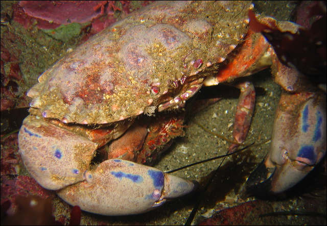
Cancer anthonyi – Yellow Rock Crab
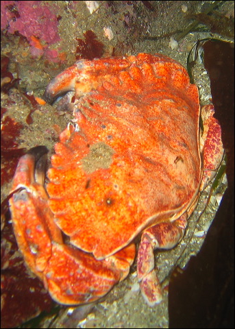
Cancer productus – Red Rock Crab
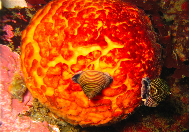
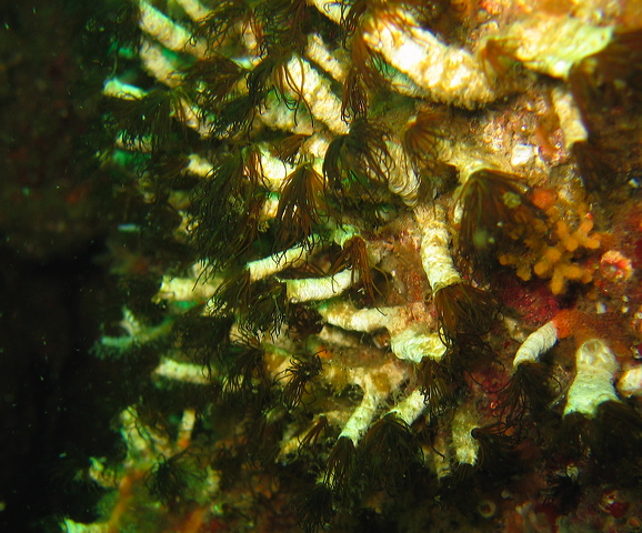
Bryozoans are colonial animals, a collaborative group of specialized individuals, called zooids. Each individual lives in a calcareous box, called a zoecium, with only the feeding tentacles clearly visible. Individuals are connected allowing food transport within the colony. Zooids are specialized, some concentrate on feeding, while others are responsible for reproduction, attachment, defense, or cleaning. Bryozoans are sessile, attached to a substrate, many are encrusting, often on kelp blades or shells. They can be confused with algae, hydroid polyps, worms, or even coral as some of the upright species build miniature reefs. They are also called moss animals, a translation of the Greek name Bryozoa, because of their plantlike appearance. They form the phylum Bryozoa, formerly called Ectoprocta. The phylum’s Greek name, Ectoprocta, means outside anus.
The body structure of the bryozoans is quite simple, arranged in bilateral symmetry without any segmentation. They have a simple nervous system without sensory organs, no complex circulatory or excretory organs. Gas exchange occurs via diffusion through the body surface. The most characteristic part of the bryozoans is the lophophore, the retractable feeding structure, a horseshoe-shaped or circular shaped set of ciliated tentacles surrounding the mouth. With the help of the lophophore bryozoans capture phytoplankton, bacteria and other microorganisms. The U shaped gut ends in an anus close to the lophophore.
Asexual reproduction, mostly budding, is responsible for the growth of the colony and the regeneration of individuals. Each colony starts from a single sexually reproduced individual, called an ancestrula. Zooids are usually hermaphroditic, eggs are often fertilized inside the colony by sperm brought in via the feeding current and brooded by the parental zooids.
Bryozoans have several predators, including sea slugs, urchins, fish. To protect themselves they secrete potent chemicals. A compound from Bugula neretina, bryostatin, demonstrates great anticancer as well as memory enhancing potentials.
Watersipora subtorquata - Invasive Japanese Bryozoan
Phidolopora labiata - Lacy Bryozoan
Heteropora pacifica – Northern Staghorn Bryozoan
Together with hydrocoral
The Greek name of the phylum, Echinodermata, means spiny skin. Instead of an external skeleton their body is supported by small calcified plates, called ossicles, and spines buried in their tissue. Most animals in this group are mobile benthic (live on the bottom) marine organisms.
They exhibit pentaradial (five-part) symmetry, where the body is arranged around an oral - aboral axis in five repeated sections. Similar to cnidarians (jellyfish and anemones), these animals perceive the world from all directions, there is no head to lead the way. However, in this case the simple radial symmetry is imposed on a much more complex body structure than that of the cnidarians. Echinoderms possess a complete gut, but no excretory organ. There is an open circulatory system but without a heart. The body fluid carries both nutrients and oxygen. As there is no head, there is no brain, their nervous system is not centralized, and sensory organs are quite limited. A unique characteristic of this group is the water vascular system, a complex series of fluid filled canals. The fleshy projections on the oral side, called tube feet or podia, often ending in suction cups, are the terminal parts of the water vascular system and are operated by water pressure. Opposite to each tube foot inside the body is a small muscular sac, the ampulla, moving the tube feet by squeezing the fluid.
Sexes are separate, fertilization usually takes place in the water column, often after synchronized spawning. The planktonic larva is bilaterally symmetrical, and only after metamorphosis obtains its pentaradial symmetry.
There are four major subgroups in this phylum with commonly seen members: sea stars, brittle stars, sea urchins, sea cucumbers. The fifth group, sea lilies and feather stars are usually found in deeper waters.
- Sea Stars
- Brittle Stars
- Sea Urchins
- Sea Cucumbers
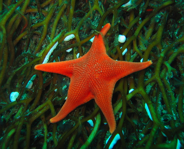
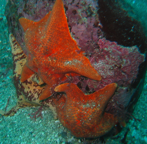
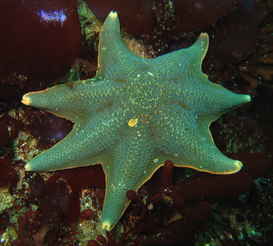
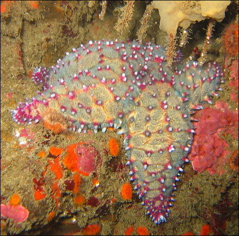
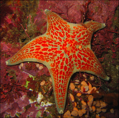
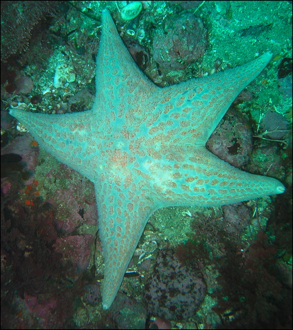
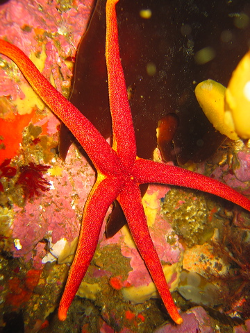
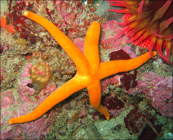
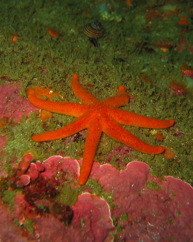
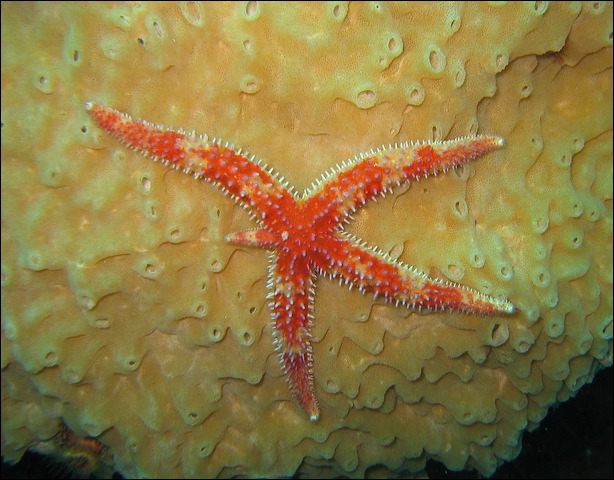
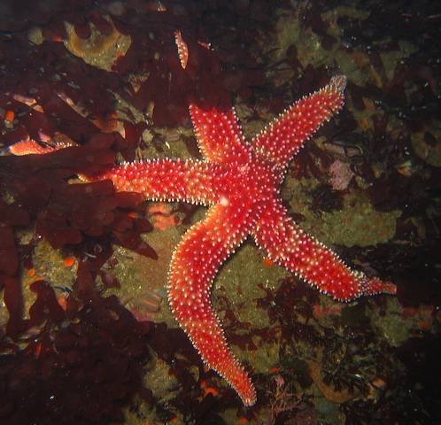
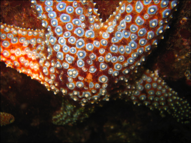
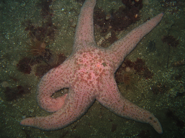
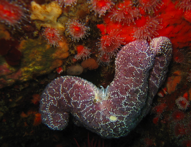
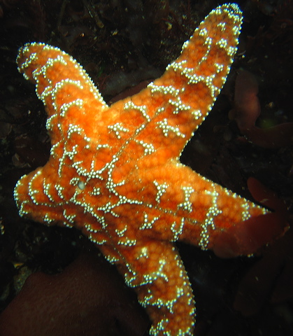
Sea Stars - Asteroidea
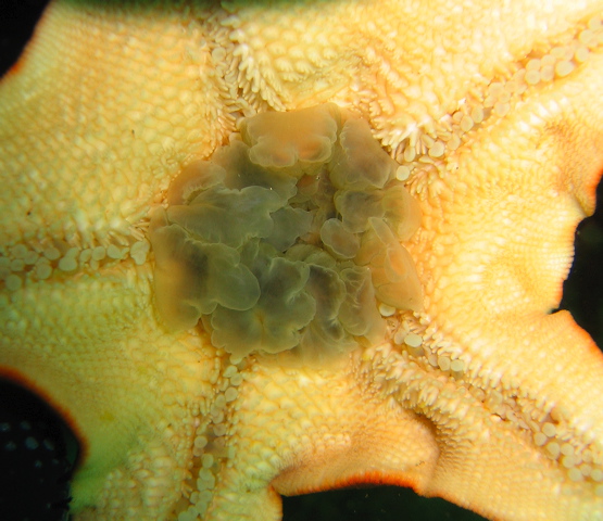
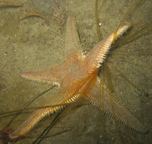
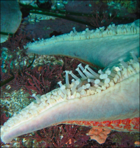
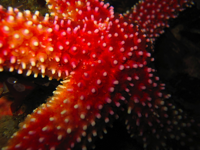
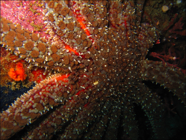
It is easy to recognize a sea star with its conspicuous star shape mostly with 5 arms. The arms, radiating from the central disk, contain internal organs, e.g. gonads and digestive glands. The mouth is in the center on the oral surface directed toward the bottom, while the anus is on the top. On the oral surface in each arm, through the ambulacral grooves, hundreds of tube feet protrude. Their function is not only locomotion and attachment, but also gas exchange, feeding, and sensory perception. On the aboral (opposite to mouth) surface a white round spot is often clearly visible. This is the madreporite, the opening to the water vascular system. To fight off predators and fouling organisms or to catch prey most sea stars use small pincers, called pedicellariae, to keep their surface clean. Gas exchange also occurs through dermal gills, protrusions of the body wall.
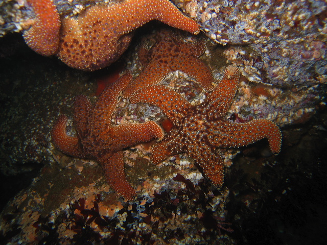
Most sea stars are carnivorous, their diet consists of snails, barnacles, mussels, anemones, some species even eat other stars, cucumbers, sponges, tunicates, algae, and detritus. Instead of swallowing their prey, most sea stars evert their stomach, envelop and digest the prey on the spot and then slurp up the digested food. The intestine is short or missing, instead there are large digestive glands. The ochre star, a key stone species, plays an important role in our intertidal area keeping the mussel populations in control. With the help of their tube feet they pull the two valves apart, inject digestive enzymes into the mussel, liquefying the prey inside its own shell. The sunflower star is a vicious predator chasing down other stars, urchins, cucumbers, crabs at impressive speed. Sea stars have many predators: otters, sealions, birds, other stars. Some species secrete foul chemicals in defense, for example the leather star has a strong garlic like smell.
Sea stars can regenerate a lost arm, and in some species an entire star can grow from a single arm if it has some of the central disk attached to it. This results in a star with one arm being much bigger than the others or a star with more or less than five arms. Some sea stars, e.g. blood star, brood their offspring, but most broadcast sperms and eggs.
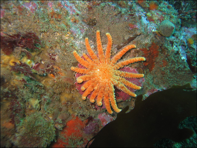
Mediaster aequalis – Red Star
Asterina miniata - Bat Star
Astropecten armatus – Spiny Sand Star
Note: Quoted northerly range is only to San Pedro, CA. Seen in Monterey, CA.
Poraniopsis inflata – Spiny Star
Dermasterias imbricata - Leather Star
Henricia aspersa - Sea Star
Henricia leviuscula - Blood Star
Solaster sp. - Sun Star
Solaster dawsoni - Dawson's Sun Star
Orthasterias koehleri – Rainbow Star
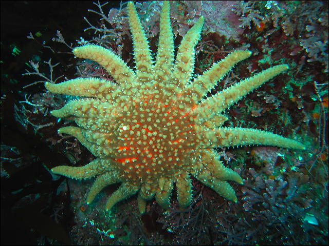
Pisaster giganteus – Giant Spined Star
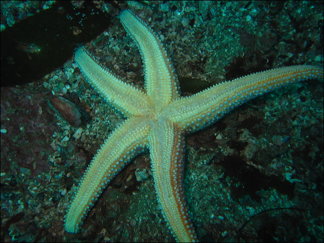
Pisaster brevispinus - Short Spined Star
Pisaster ochraceus – Ochre Star
Pteraster tesselatus – Cushion Star
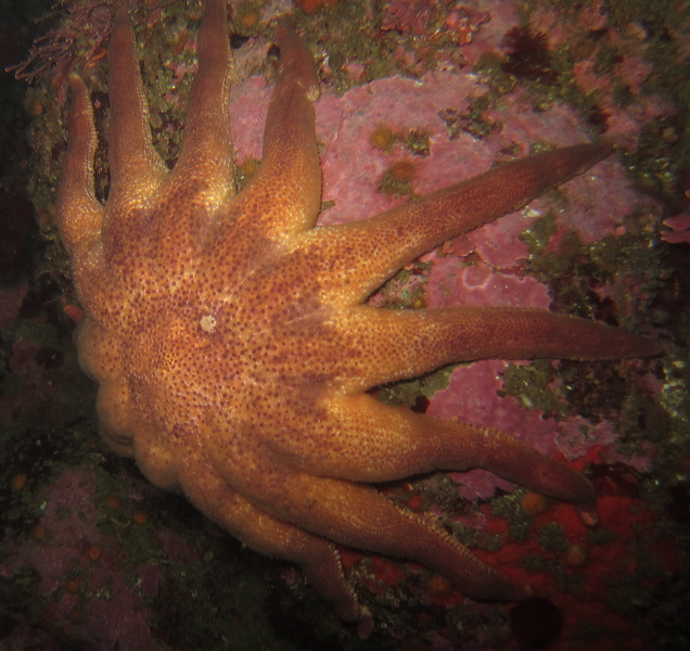
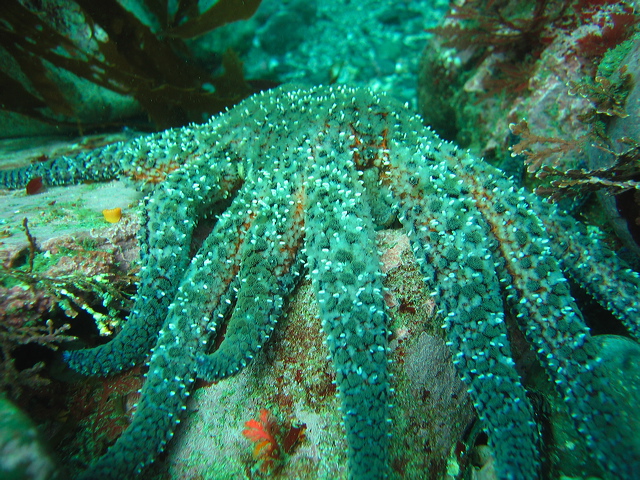
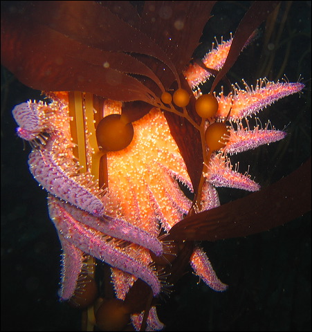
Pycnopodia helianthoides – Sunflower Star
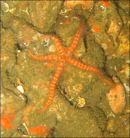
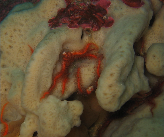
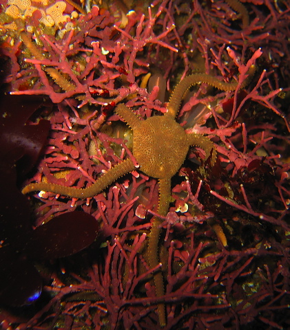
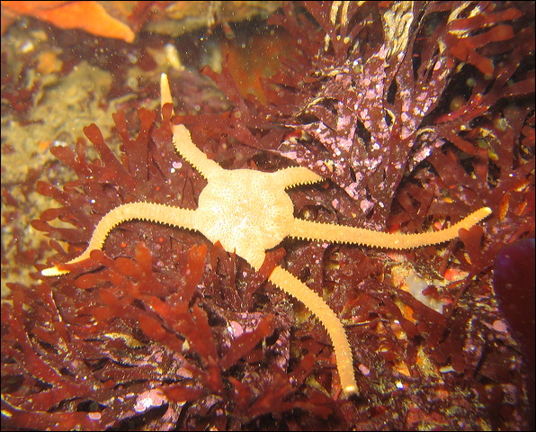
Brittle stars - Ophiuroidea
Brittle stars have a similar star shaped body, but the central disk is much smaller than in sea stars and the arms are relatively long and flexible. The tube feet do not have suckers and are used for feeding, sensing light or chemicals, and burrowing. Locomotion is achieved mainly by a swimming motion of the arms. In contrast to sea stars, all organs are contained in the central disk and they lack an anus. Brittle stars are so called because they readily dislodge an arm, which is easily regenerated. Brittle stars look like worms to the untrained eye, often found hidden in the kelp holdfast or in large sponges or tunicates by the hundreds. Brittle stars are both suspension and deposit feeders often using mucus in capturing food. They are preyed upon by perches and greenlings.
Ophiothrix spiculata – Spiny Brittle Star
Ophioplocus esmarki - Smooth Brittle Star
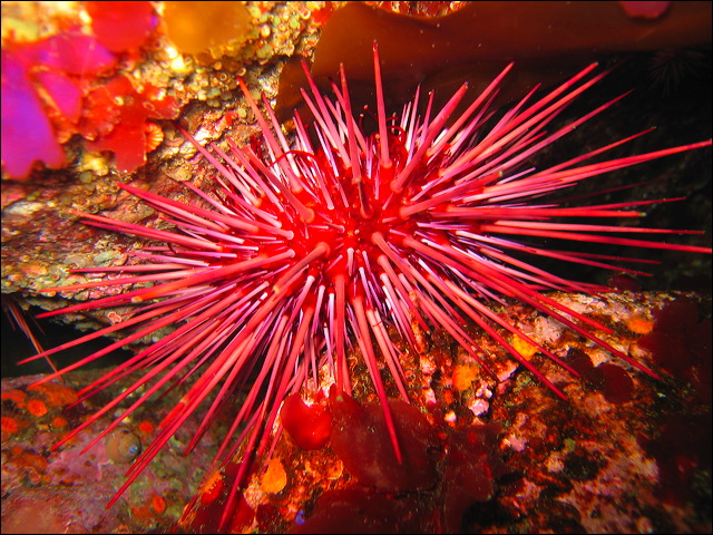
Sea urchins - Echinoidea
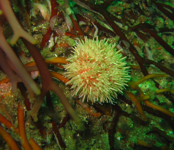
Based on casual comparison it is not obvious that sea urchins and sea stars are related. The endoskeleton consists of a spherical rigid test and flexible spines covered with a thin tissue layer. Imagine the arms folded upwards so the five rows of ambulacral grooves with tube feet are located on the top side of the sphere. Urchins are herbivores, their main diet is seaweed, especially giant kelp around this area. An out of control urchin population can cause urchin barrens, a total destruction of the kelp forest. Small pieces of kelp are passed down by the spines and tube feet to the mouth located at the bottom. In the mouth there is a complicated jaw structure, called Aristotle’s lantern, composed of hard plates and muscles controlling the movement of five teeth. It is strong enough to carve depressions into rocks and shale. Sea otters and sheepheads are the main predators of urchins around here, but you can find urchin also in sushi restaurants as uni.
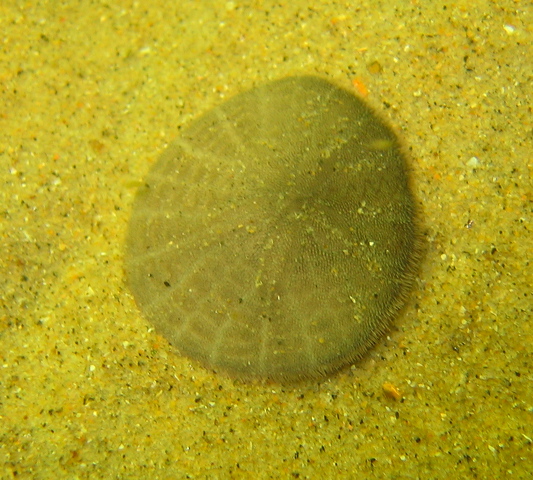
Heart urchins and sand dollars are also in this group, their body is flattened evolved for living in soft bottom
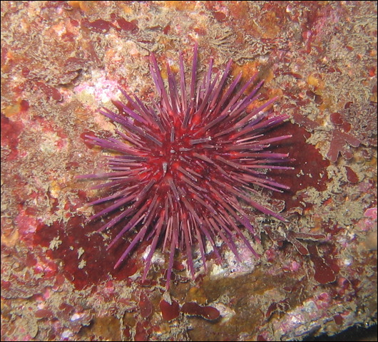
Strongylocentrotus franciscanus - Red Sea Urchin
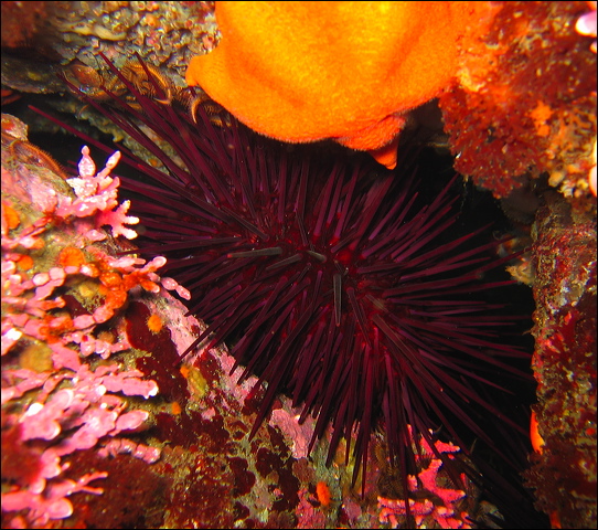
Strongylocentrotus purpuratus - Purple Sea Urchin
Dendraster excentricus – Sand Dollar
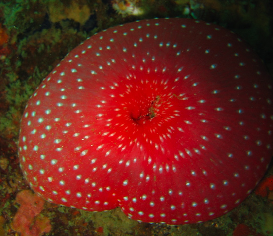
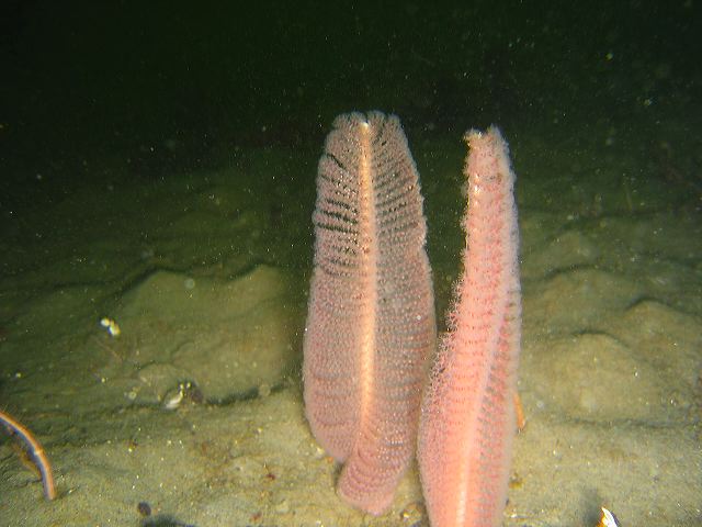
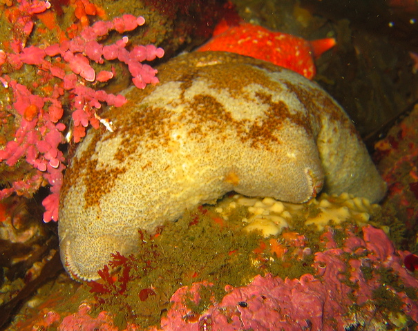
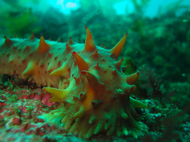
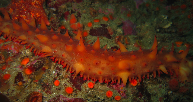
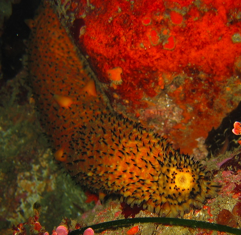
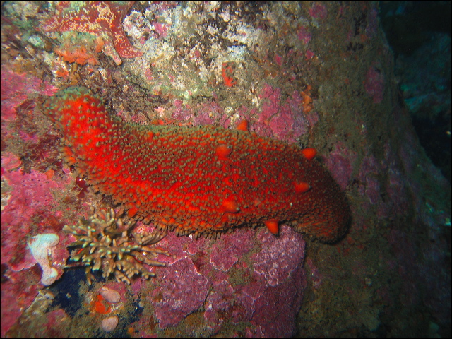
Sea cucumbers – Holothuroidea
The sausage like body of a sea cucumber seems to be related to slugs rather than sea stars and urchins. Their body is elongated along the oral – aboral axis, mouth and anus are far apart. The five rows of tube feet are along the bottom side. The skeletal pieces are small and far apart, hence the soft, squishy appearance. Cucumbers can easily change their body shape from sausage to football. Under stress as a defensive act some cucumbers eviscerate their gut and other internal organs, which can be regenerated in a few weeks. Close to the anus is the respiratory tree, a unique structure of branched tubes performing gas exchange through its large surface as water is pumped in from the anus.
Cucumbers are deposit or suspension feeders, they are the vacuum cleaners of the ocean. The tube feet close to the mouth have evolved into tentacles to pick up sediment and detritus from the bottom. Their intestine is especially long for absorbing organic matter from sediments. Some species, like the orange cucumber capture food directly from the water column using their large tentacles extended between the rocks. Sea stars, sea otters, even humans eat sea cucumbers.
Cucumaria piperata – Peppered Sea Cucumber
Cucumaria miniata – Orange Sea Cucumber
Parastichopus californicus – California Sea Cucumber
Parastichopus parvimensis – Warty Sea Cucumber
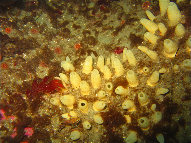
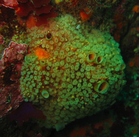
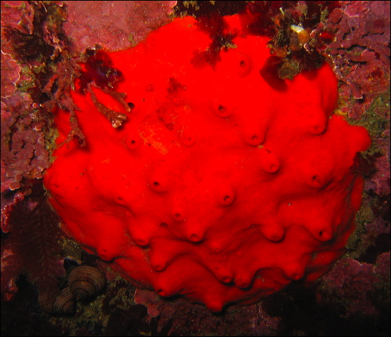
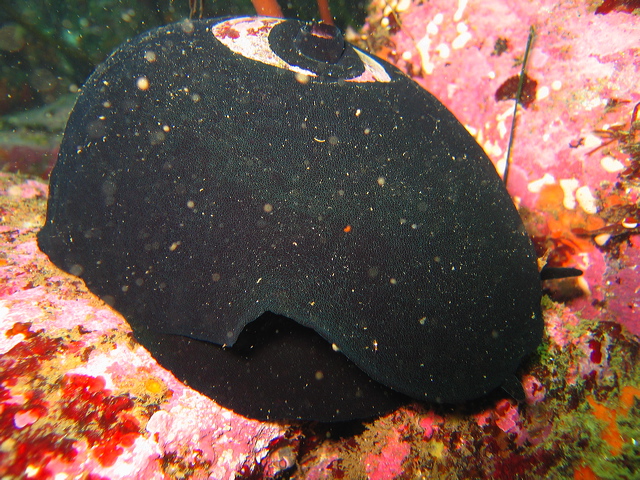
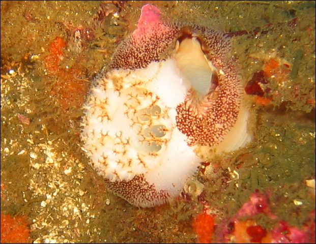
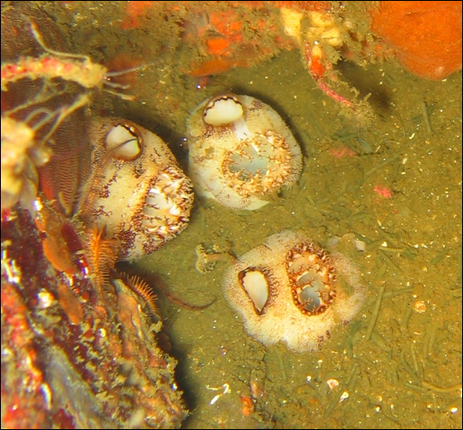
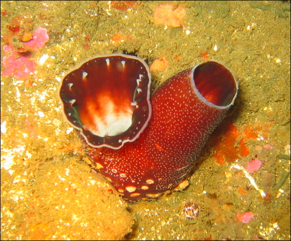
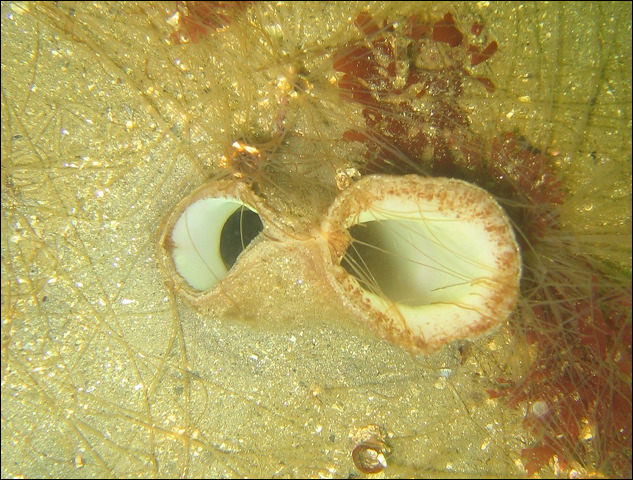
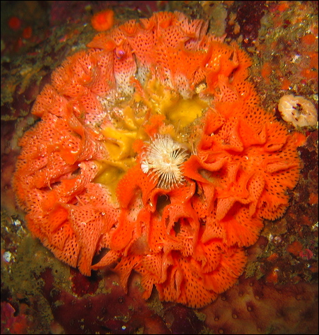
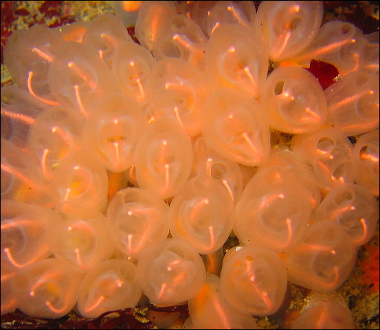
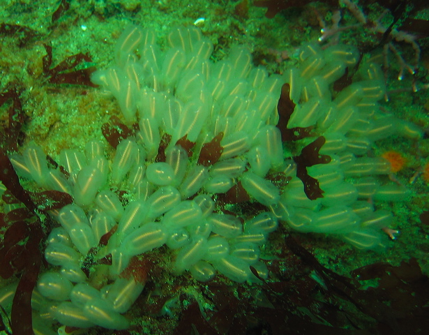
This group of animals is a subphylum in the Phylum Chordata and as such they are our closest invertebrate relatives. Tunicates are chordates without a backbone, however, they possess the chordate characteristics (notochord, dorsal hollow nerve cord, pharyngeal gill slits, post anal tail) only in their larval stage, the adults we encounter couldn’t differ more from us. The ascidians, the class Ascidiacea, are benthic sessile tunicates, attached to hard substrates, in contrast to the pelagic tunicates or salps which are not covered here. Ascidian tunicates come in three main forms. Solitary tunicates are relatively large single individuals. The social tunicates live as a group of individuals connected to each other. The compound tunicates are composed of many small individual zooids living in a common gelatinous matrix. The body is covered with a gelatinous tunic made of cellulose, a material almost exclusive to plants. Compound tunicates are easily confused with sponges, but there are some clues to separate them. Sponges have a felty appearance, while compound tunicates usually have a shiny surface. The incurrent openings are usually not visible in sponges, so regularly placed small pores are characteristics of the tunicates.
Although they are similar in appearance, the tunicate body structure is much more complex than the simple cell aggregation of sponges. Similar to sponges, tunicates are filter feeders. Water enters the body at the incurrent, oral siphon, passes through the mucus covered pharyngeal basket where small particles are filtered out. Food then passes through the U shaped gut for digestion and the water is expelled through the excurrent, atrial siphon. Many other groups of invertebrates rely on creating water current for filter feeding, but using a perforated pharinx to catch particles is a highly evolved mechanism and is unique to tunicates. The water current is maintained by hundreds of beating cilia, but it can be regulated also by muscles. If disturbed, tunicates squirt water out through both siphons, hence the common name, sea squirt. Blood is pumped around the body by a short tubular heart. For approximately 100 beats the blood is flowing in one direction, then after a short pause it starts flowing the opposite direction. The tunicate blood has a very high content of heavy metals, especially vanadium and iron, possibly to deter predators. Gas exchange occurs across the body wall. There is no head, the nervous system and sensory organs are not highly developed, but adequate for the sessile life style.
Most tunicates are hermaphroditic with separate gonads. Most solitary tunicates broadcast sperms and eggs, the external fertilization produces a tadpole larva which displays more chordate features than the adults. In contrast, the compound tunicates brood their internally fertilized eggs. In social and compound tunicates asexual budding is responsible for the fast growth of the colony.
To deter predators, similar to sponges, tunicates secrete various chemicals that are of pharmacological interest. Yondelis, a recently approved drug, was developed from a tunicate to treat soft tissue sarcoma. Due to this chemical defense system tunicates do not have many predators.
Solitary tunicates
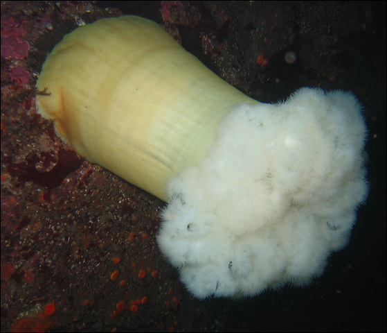
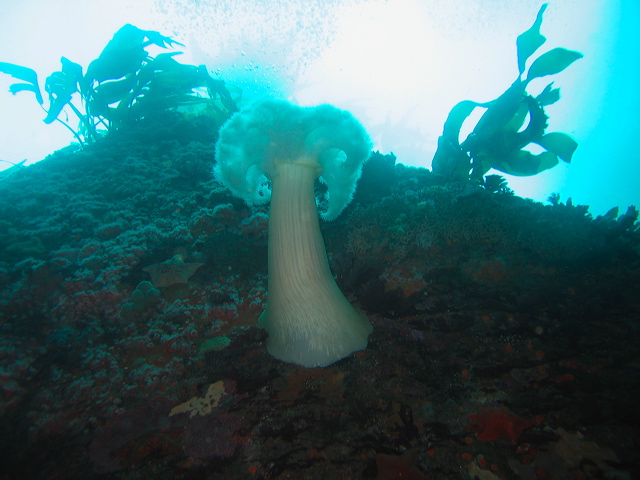
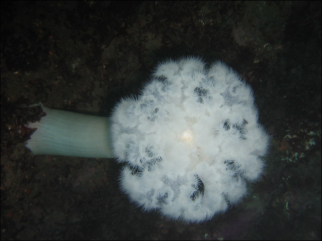
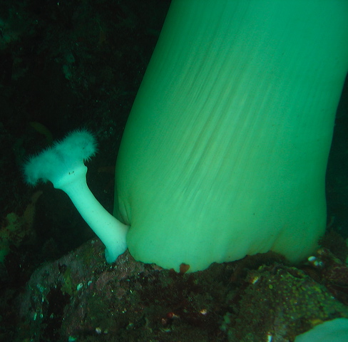
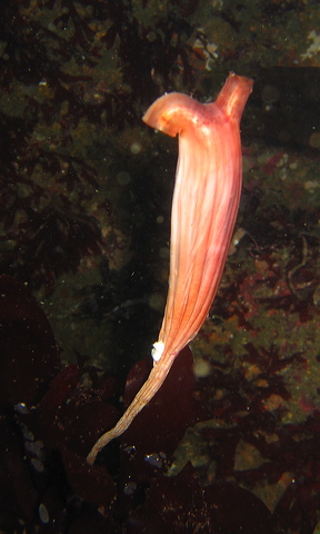
Styela montereyensis – Stalked Tunicate
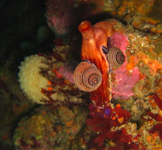
With snails
Cnemidocarpa finmarkiensis – Solitary Tunicate
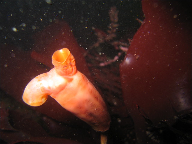
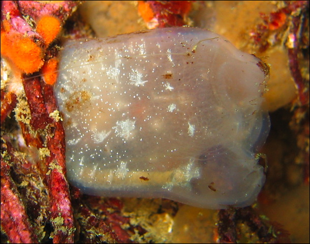
Corella willmeriana - Solitary Tunicate
ID ??
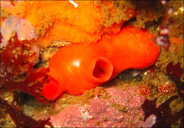
Boltenia villosa - Spiny Headed Tunicate
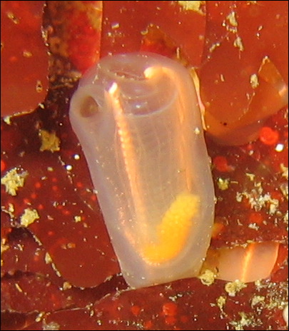
Ascidia paratropa - Glassy Tunicate
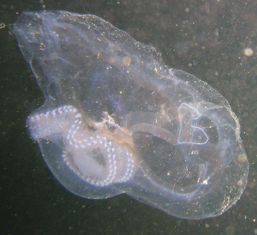
Family: Salpidae
Cyclosalpa sp? (The specimen is incomplete)
Social tunicates
Clavelina huntsmani – Lightbulb Tunicate
Euherdmania claviformis
Metandrocarpa taylori - Colonial Tunicate
Compound tunicate
Cystodytes lobatus – Lobed Tunicate
Didemnum carnulentum
Diplosoma macdonaldi
Aplidium californicum
close up
Aplidium sp.
Archidistoma molle
Updated: 7 July 2012